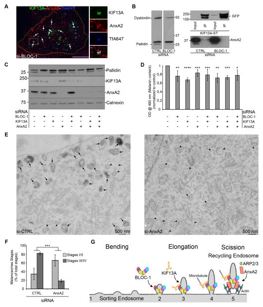
Figure 6. Annexin A2, BLOC-1 and KIF13A cooperate for melanosomes biogenesis (related to Supplemental Figure S6).
(A) IFM of AnxA2 (red) on KIF13A (green)-expressing siBLOC-1 HeLa cells that internalized TfA647 (blue). Arrows, triple KIF13A, AnxA2 and Tf+ endosomes. (B) WB representative of 3 independent IPs of GFP-KIF13A-ST expressing siCTRL or siBLOC-1 HeLa cells lysates (left) analyzed using GFP (top right) or AnxA2 (bottom right) antibodies. Lower AnxA2 amount is revealed in siBLOC-1 IP while greater KIF13A-ST was immunoprecipitated relative to controls. (C) WB of siRNAs treated MNT1 lysates using pallidin (top), KIF13A (middle top), AnxA2 (middle bottom) or calnexin (bottom) antibodies. (D) Intracellular melanin quantification of MNT1 cells as in C. (E) Conventional EM analysis of siCTRL or siAnxA2 MNT1 cells. Arrowheads, immature melanosomes stages I/II; arrows, mature pigmented melanosomes stages III/IV. (F) Quantification of melanosomes stages in siCTRL or siAnxA2 MNT1 cells. (G) Model of recycling endosomes shaping by BLOC-1. BLOC-1 binds to highly curved SE membrane deformations (1–2). KIF13A interacts with BLOC-1 at the newly formed tubule (3) and generates the pulling force to elongate the RE tubule from SE (4). AnxA2 binds to SE elongated bud, promotes the ARP2/3-dependent polymerization of branched actin filaments and participates to the stabilization and/or scission of RE tubules (5). Data represent the average of at least 3 independent experiments, normalized to control and presented as a mean ± SD. Scale bars: 500 nm (EM); 10 μm (IFM). ****, p<0.001; ***, p<0.01; **, p<0.02; *, p<0.05.

