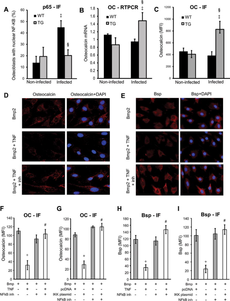
Fig. 1.

NF-κB-mediated response inhibits bone and matrix protein synthesis. Destructive periodontitis was initiated in IKK-DN transgenic mice (TG) or wild-type (WT) control mice by oral inoculation of the periodontal pathogens P. gingivalis plus F. nucleatum and compared with vehicle alone. Mice were euthanized 6 weeks after oral inoculation. Nuclear NF-κB was measured by immunofluorescence by colocalization of p65, a subunit of NF-κB and DAPI nuclear staining. (A) Number of NF-κB-positive cells was determined. Localization to bone-lining osteoblastic cells was determined in cells with a cuboidal appearance and with a nucleus that was not fusiform or parallel to the bone surface. (B) RNA was isolated from periodontal tissue, and osteocalcin mRNA levels were measured by real-time PCR. (C) OC expression was measured by immunofluorescence with antibody to OC and expressed as the mean fluorescence intensity. (D–I) MC3T3 cells were incubated with TNFα or transfected with IKK-overexpression vector compared with pcDNA (empty vector) alone. NF-κB was inhibited with the specific inhibitor BAY117082. Cells were stimulated with Bmp2 for 48 hours. Cells were stained for OC or Bsp (red) and counterstained with DAPI (blue). Immunofluorescence was analyzed and expressed as mean expression above baseline (MFI). ‡Significantly different in infected compared with matched noninfected group; §significantly different in infected TG compared with infected WT. Data shown are representative of three independent experiments. +Significant inhibition with TNFα or IKK transfection; #significantly different between cells incubated with no inhibitor.
