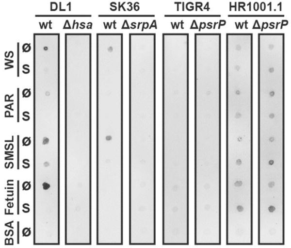
Fig. 1.

Streptococcal adhesion to immobilized whole saliva, parotid saliva and submandibular/sublingual saliva. Suspensions of fluorochrome-labeled S. gordonii DL1, S. sanguinis SK36, S. pneumoniae wild-type strain TIGR4, capsule-free mutant strain HR1001.1, and the respective isogenic SRR adhesin-deficient mutants were overlaid on nitrocellulose membranes carrying immobilized untreated (Ø) or sialidase-treated (S) dots (1 μg of total protein per dot) of whole saliva (WS), parotid saliva (PAR), and submandibular/sublingual saliva (SMSL). Fetuin and bovine serum albumin (BSA) were included as controls. Fluorescent signals of bound bacteria were detected by use of a fluorescence scanner.
