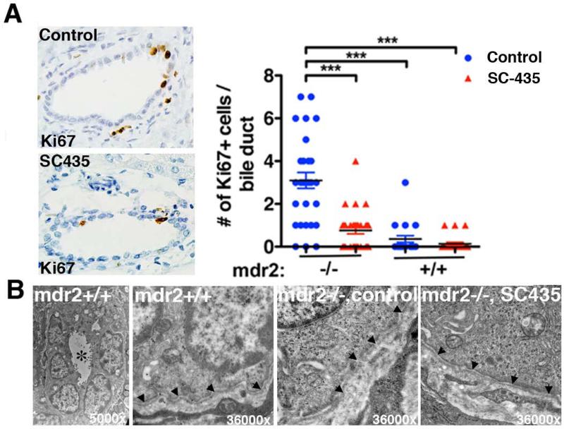
Figure 6. SC-435 treatment is associated with decreased bile duct epithelial injury and partial restoration of ultrastructural integrity.
Paraffin-embedded liver sections from SC-435 treated and control mice (n=2-3/group) were subjected to Ki67 immunohistochemistry. Ki67 positive cells were enumerated in large interlobular bile ducts (10 bile duct profiles/ liver). Statistical analysis: one-way ANOVA was applied to compare the data between the groups (A). In B, glutaraldehyde-fixed liver sections were analyzed by electron microscopy. Left panel depicts a bile duct profile in control mdr2+/+ mice in low magnification, with * denoting the lumen. The other images show higher magnification of the basement membrane (denoted by arrowheads) in mdr2+/+ mice, which is disrupted in control mdr2−/− mice; integrity is restored in SC-435-treated mdr2−/− mice. 10 bile duct profiles were examined per group (n=2 mice/group), of which 4 were surrounded by intact basement membrane in control and 7 in SC-435 treated mice.

