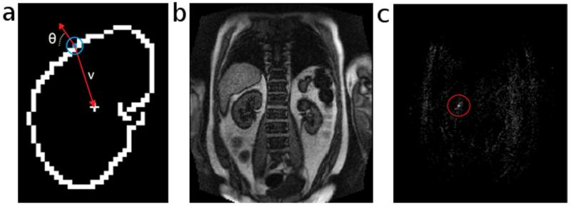
Figure 2.
Identifying the center of the kidney with the generalized Hough transform. a) Template of a kidney contour. Each edge pixel (colored blue in the blue circle) along the contour is indexed by its orientation angle (θ) and a vector (v) that points towards the contour’s center. b) An MRR image frame that we want to register to the template. c) A map with the same matrix size as the image frame. The value of each element represents the number of edge pixels that regard the current element as the center of the kidney contour in the current frame. The brightest point in the red circle is the true center, and is used for registering the current frame to the template.

