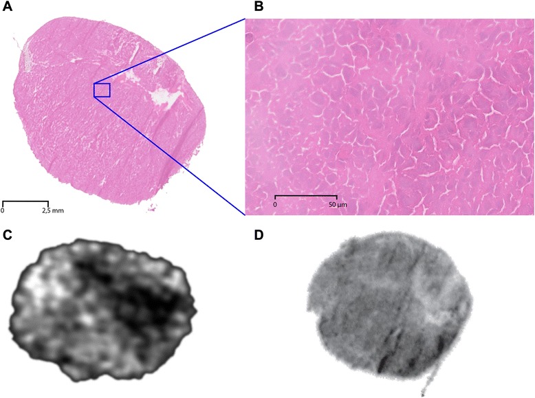
Fig. 1.

H&E staining of a PC295 tumor showing homogenous tissue density (a) and viable tissue as seen on an enlarged section (b). Single tumor slice from SPECT image demonstrating heterogeneous peptide distribution in vivo (c) despite high and homogeneous receptor density throughout the tumor as assessed by in vitro autoradiography (d)
