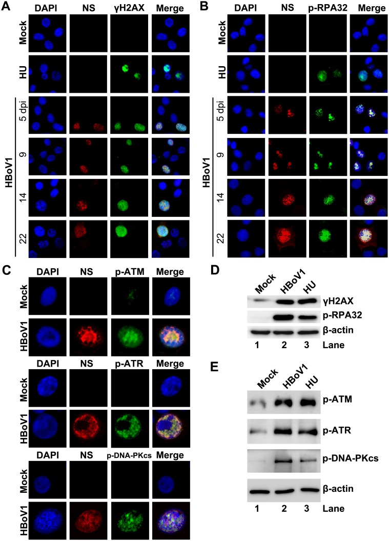
Fig 2. HBoV1 infection of HAE-ALI cultures induces phosphorylation of H2AX and RPA32 and activates ATM, ATR, and DNA-PKcs.
HAE-ALI cultures were infected with HBoV1 or mock-infected. (A, B, and C) IF analysis. (A and B) At the indicated dpi, the infected cells trypsinized off the insert were cytospun and co-immunostained with anti-NS1C and anti-γH2AX (A), and with anti-NS1C and anti-p-RPA32 (B). (C) At 10 dpi, infected cells trypsinized off the ALI membrane were used for IF analysis with anti-NS1C and p-ATM, with anti-NS1C and anti-p-ATR, and with anti-NS1C and anti-p-DNA-PKcs, as indicated. Nuclei were stained with DAPI (blue), and the cells were visualized by confocal microscopy at a magnification of × 100. (D and E) Western-blot analysis. At 10 dpi, cells of the mock-, HBoV1-infected, or HU-treated HAE-ALI cultures were lysed in 1 × SDS-containing loading buffer. Equivalent volumes of the lysate were used for Western blot using anti-γH2AX, and reprobed with p-RPA32 and anti-β-actin, sequentially (D), and using with anti-p-ATM, anti-p-ATR, anti-p-DNA-PKcs, and anti-β-actin, respectively (E). HAE-ALI cultures treated with HU at a final concentration of 2 mM for 2 days were used as positive control.

