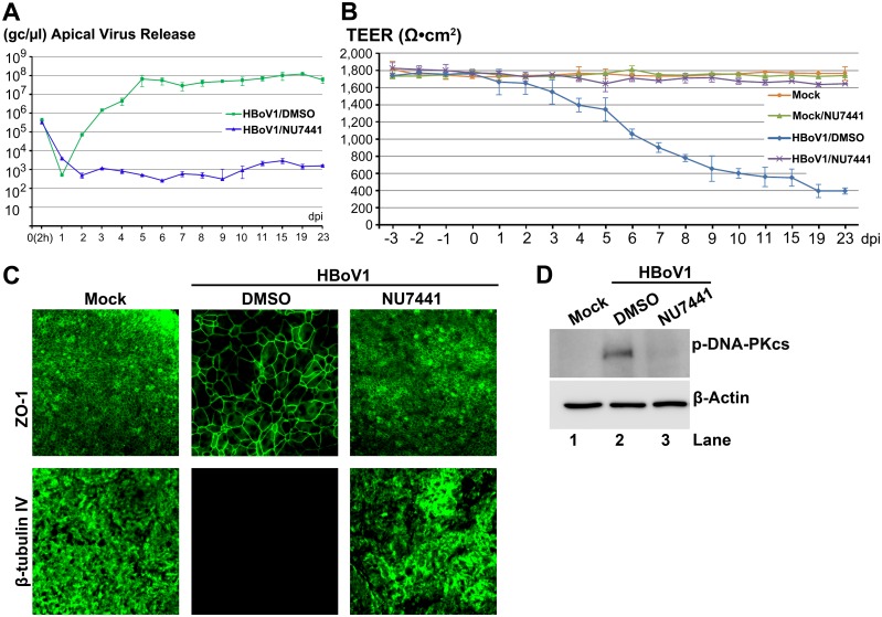
Fig 5. A DNA-PKcs-specific inhibitor decreases HBoV1 infection of HAE-ALI.
At two days prior to apical infection of HBoV1, HAE-ALI cultures were incubated with NU7441 at 20 μM in the basolateral chamber. (A) Quantification of apical virus release. At the indicated dpi, apical washes were quantified for HBoV1 genome copies qPCR (Y-axis) and plotted to the dpi as shown. Means and standard deviations (n = 3) are shown. (B) TEER measurement. At the indicated dpi, the TEER of infected HAE-ALI cultures, as indicated, was determined. Means and standard deviations (n = 3) are shown. (C) IF analysis. At 23 dpi, the ALI membrane of the infected HAE-ALI cultures was stained with anti-β-tubulin IV or with anti-ZO-1, as indicated. The stained membranes were visualized for β-tubulin IV/ZO-1 (green) expression by confocal microscopy at a magnification of × 40. (D) Analysis of phosphorylated DNA-PKcs. At 23 dpi, equivalent cells of the infected HAE-ALI cultures were analyzed by Western blotting for expression of p-DNA-PKcs and β-actin, respectively.

