Abstract
Sake is a traditional Japanese alcoholic beverage that is gaining popularity worldwide. Although sake is reported to have beneficial health effects, it is not known whether chronic sake consumption modulates health risks due to radiation exposure or other factors. Here, the effects of chronic administration of sake on radiation-induced metabolic alterations in the livers of mice were evaluated. Sake (junmai-shu) was administered daily to female mice (C3H/He) for one month, and the mice were exposed to fractionated doses of X-rays (0.75 Gy/day) for the last four days of the sake administration period. For comparative analysis, a group of mice were administered 15% (v/v) ethanol in water instead of sake. Metabolites in the liver were analyzed by capillary electrophoresis-time-of-flight mass spectrometry one day following the last exposure to radiation. The metabolite profiles of mice chronically administered sake in combination with radiation showed marked changes in purine, pyrimidine, and glutathione (GSH) metabolism, which were only partially altered by radiation or sake administration alone. Notably, the changes in GSH metabolism were not observed in mice treated with radiation following chronic administration of 15% ethanol in water. Changes in several metabolites, including methionine and valine, were induced by radiation alone, but were not detected in the livers of mice who received chronic administration of sake. In addition, the chronic administration of sake increased the level of serum triglycerides, although radiation exposure suppressed this increase. Taken together, the present findings suggest that chronic sake consumption promotes GSH metabolism and anti-oxidative activities in the liver, and thereby may contribute to minimizing the adverse effects associated with radiation.
Introduction
Human health is influenced by lifestyle choices, including exercise, diet, and tobacco use. Among these factors, alcohol consumption is related with numerous health risks and diseases, such as cardiovascular diseases and cancer [1]. The liver is particularly susceptible to alcohol-related disease [2] and higher alcohol consumption is a major cause of hepatocellular carcinoma [3]. In addition, when combined with other risk factors, such as tobacco, alcohol may have synergistic effects on health [4–7].
Radiation is also a significant health risk factor, and is associated with both acute and chronic effects depending on the quality and dose of radiation. The effects of radiation on human health may be mediated, at least in part, by lifestyle-related factors, such as diet [8–10]. For example, the findings from epidemiological studies in nuclear plant workers suggest that lifestyle-related factors, including alcohol consumption, influence the adverse effects of radiation [11,12]. However, how alcohol drinking custom mediates radiation effects remains unclear.
Many types of alcohol beverages are consumed worldwide. Although alcohol in general has been shown to adversely affect human health [2,13], certain beverages, including beer and sake, have been demonstrated to have anti-mutagenic activities [14]. Recently, it was reported that sake, a popular Japanese alcoholic beverage, has protective effects against acute radiation [15]. However, it remains unknown whether chronic sake consumption mediates such radiation-induced effects.
The liver is the main organ involved in detoxification of harmful substances, including alcohol [16] and is susceptible to radiation damage [8,10,17]. Alcohol is metabolized in the liver, and the resulting metabolic byproducts can impair liver function and cause tissue damage [18]. For this reason, liver metabolites are useful indicators of health status.
Here, the influence of chronic sake consumption on radiation-induced effects, particularly those related to the alteration of liver metabolites, was evaluated in mice using a metabolic approach.
Materials and Methods
Animal care
Seven week-old female C3H/He mice were purchased from the Japan SLC Co. (Hamamatsu, Japan). Mice were housed for seven weeks to allow for adaptation before performing experiments. Mice were typically allowed access to water and standard laboratory chow (MB-1, Funabashi Farm Co., Japan) ad libitum. The major components of MB-1 (gross energy, 4.28 Kcal/g) were as follows: total carbohydrate, 54.4%; proteins, 24.2%; fat, 4.4%; fibers, 3.6%; moisture, 8.0% and ash, 5.4%. All animal studies were reviewed and approved by The Institutional Animal Care and Use Committee of the National Institute of Radiological Sciences (NIRS), and were performed in strict accordance with the NIRS Guidelines for the Care and Use of Laboratory Animals. A total of 4 or 5 mice were used in each administration group (sake or ethanol). Measurements of whole body weight, organ weight, and metabolic markers were performed using 3 (corresponding to metabolome analysis samples) or 4 mice in each administration group in two independent experiments.
Sake administration
Sake (junmai-shu; Daishichi Sake Brewery, Nihonmatsu, Japan) produced with rice polished to 69% and containing 15% (v/v) alcohol was used to examine the effects of sake on the liver metabolome in this study. As a comparative study to examine the effects of ethanol, 15% (v/v) special grade ethanol (99.5%) (Wako Pure Chemical Industries, Ltd.) in water was administered to mice instead of sake.
Sake (0.2 or 0.6 ml per 23 g body weight, corresponding to 0.009 ml/g or 0.026 ml/g body weight, respectively) was administrated to mice using a feeding needle every morning for one month (30–31 days). The body weights of mice were measured every evening and used for calculation of the sake intake amounts the next morning. In comparative experiments using 15% ethanol in water, 0.6 ml of ethanol solution was administered to mice. A control group was administered the same volume of drinking water in place of sake. Before administering the test solutions in the morning, food and water were withheld from mice from the previous evening. Mice in the irradiated group were irradiated on the last four days of the sake-administration period, as described in the following section. A total of 4 or 5 mice were used in each experimental group for sake and ethanol. S1 and S2 Figs are representative graphs showing changes in the body weights of mice administered either sake or 15% ethanol administration. After giving the mice 0.6 ml sake per day for one month, the gross appearance of mice was normal and no changes in weight were observed compared to controls; therefore, the following experiments were performed by administering 0.6 ml sake to mice, unless otherwise noted.
Irradiation
Mice orally administered sake every day for one month (0.6 ml per 23 g body weight) were treated with fractionated irradiation (0.75 Gy/day). The irradiation was performed once a day at a dose rate of 0.85 Gy/min during the last four days of the sake- or ethanol- administration period immediately after the administration. Irradiation was performed using a Pantak 320S machine (Shimadzu, Japan) equipped with a 0.50-mm Al + 0.50-mm Cu filter and operated at 200 kVp and 20 mA. An exposure rate meter (AE-1321M; Applied Engineering Inc., Japan) was used for the dosimetry. Blood and organ collection was performed one day after the last irradiation. Mice were anesthetized by inhalation of gaseous isoflurane (Pfizer, Tokyo, Japan) and blood was collected for serum preparation. The mice were then euthanized by cervical dislocation, and liver, thymus and spleen samples were collected for analysis.
Metabolome analysis
The liver tissue from three mice from the sake and ethanol administration groups was used for metabolome analysis. A portion of the collected liver tissue was immediately frozen in liquid nitrogen for metabolite analysis. After thawing, the samples (approximately 50 mg) were homogenized in 1800 μl of 50% acetonitrile solution (v/v) containing internal standards (Human Metabolome Technologies (HMT), Inc., Tsuruoka, Japan) at concentrations of 5 and 20 μM for anion and cation modes, respectively. The homogenized samples were centrifuged (2,300 × g) for 5 min at 4°C and the separated upper layer was ultrafiltered (9,100 × g for 120 min at 4°C) using an ultrafiltration tube with a molecular weight cut-off of 5 kDa. The filtrate was evaporated and dissolved in 50 μl Milli-Q water for analysis using a capillary electrophoresis-time-of-flight mass spectrometry (CE-TOFMS) system (Agilent Technologies). Cationic metabolites were diluted twofold in cation buffer solution (HMT, Inc.) and analyzed using a fused silica capillary tube (i.d. 50 μm × 80 cm). For the analysis, the samples were injected into the system at a pressure of 50 mbar over 10 sec. The CE voltage was set at 27 kV. Electro-spray ionization-mass spectrometry (ESI-MS) was conducted in the positive ion mode. The capillary voltage was set at 4000 V. The spectrometer was scanned from m/z 50–1000. Anionic metabolites were diluted fivefold in anion buffer solution (HMT, Inc.) and analyzed using a fused silica capillary tube (i.d. 50 μm × 80 cm). For the analysis, the samples were injected at a pressure of 50 mbar over 25 sec. The CE voltage was set at 30 kV. ESI-MS was conducted in the negative ion mode. The capillary voltage was set at 3500 V. The spectrometer was scanned from m/z 50–1000. The raw data obtained by CE-TOFMS were processed with MasterHands software (ver. 2.16.0.15; developed at Keio University, Japan), which automatically extracted peaks greater than 3 (Signal/Noise ratio), and various data, including the m/z ratio, migration times (MT), and peak area values, were collected. The relative area values were calculated based on the sample tissue weight and were adjusted for the analysis sensitivity. Each peak was aligned based on the MT and m/z value. Metabolites in the samples were identified by comparing the MT and m/z values with those of authentic standards (HMT metabolite library), in which differences of ± 0.5 min and ± 10 ppm were permitted. The estimated relative area values were subjected to principal component analysis (PCA). We used the technical services of the HMT Research Group (Tsuruoka, Japan) for the metabolome analysis by CE-TOFMS. PCA was performed using SampleStat ver.3.14 (HMT). Hierarchical clustering analysis (HCA) and heat mapping were performed using PeakStat ver3.18 (HMT). The identification of metabolites was performed using the HMT database by measuring standard metabolites. This method for identification has been used in many organisms and it is also accepted in liver samples [19, 20, 21]. Description of XA・・・or XC・・・ in the compound name in S3 and S4 Tables indicate unknown peaks that have been detected in samples from other organisms in the HMT database for metabolite identification.
Free amino acid analysis
Free amino acid concentrations in sake were determined using an automated amino acid analysis system (JLC-500v2; JEOL Ltd., Japan). The assay samples were prepared by adding 22 ml of 0.1% 2-mercaptoethanol and 3 ml of 50% TCA solution to each 5-g sample of sake. After mixing, the resulting solutions were kept for 3 hours on ice, and were then centrifuged at 10,000 × g for 20 min at 4°C. After filtration of the supernatants through a No. 5A filter (Advantec), 1 N NaOH (70 μl) was added to 1 ml of filtrate, and the resulting solutions were diluted 1:3 (v/v) in the primary buffer of the analysis system. After filtration through a 0.45μm filter (DISMIC-13CP, Advantec), the samples were analyzed. All procedures were performed on ice. All analyses were performed by the NH (Nipponham) Foods Ltd. Research and Development Center (Tsukuba, Japan).
Serum preparation and biochemical marker analysis
Collected blood was kept at room temperature for 90 min and was then centrifuged at 1,000 × g for 15 min at 20°C. The resulting supernatant was collected as serum and was stored at -80°C until needed for analysis. Metabolic markers in serum were analyzed using a Dri-Chem 7000V (Fuji Film, Japan).
Statistical analysis
Changes in metabolites were examined statistically using Welch’s t-test. To draw biological inferences using factor loading in the principal component analysis (PCA), factor loading was defined as the correlation coefficient between the PC scores and variables [22]. Statistical testing for factor loading in PCA was performed based on the fact that for a correlation coefficient r, the statistic:
has a t-distribution with (n−2) degrees of freedom. Metabolites that has statistically significant (P<0.01) correlation between the PC score and the relative area value were selected, and characters of the metabolites were evaluated. All other statistical analyses were performed by the unpaired t-test.
Results and Discussion
Effects of sake and radiation on organ weight
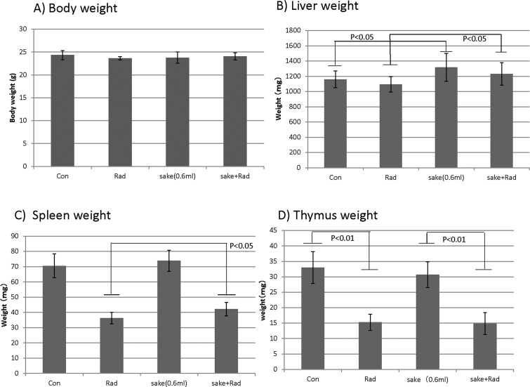
Mice administered pure Japanese sake (junmai-shu) or 15% (v/v) ethanol in water for one month appeared normal. During the one-month administration period, significant decreases in the body weights of mice in the groups administered ethanol or sake compared to the no intake group were observed; however, no significant difference in mean body weight was detectable at the end of the administration period compared to the control group. (S1 and S2 Figs and S1 and S2 Tables). Body and organ weights (liver, thymus, and spleen) of the mice administered with sake were also evaluated when blood and liver tissues were collected. No significant differences in body weights were observed between any of the treatment conditions (Fig 1A). However, liver weights increased slightly but significantly in mice administered sake, although radiation had no marked effects on liver weight (Fig 1B). The observed decreases in spleen weights in irradiated mice were slightly but significantly reversed by the administration of sake (Fig 1C). For the thymus, the marked decreases in weight induced by irradiation were similarly observed in irradiated mice administered sake (Fig 1D).
Fig 1. Effects of sake on the weight of the body, liver, spleen and thymus of irradiated mice.
The weights of the (A) body, (B) liver, (C) spleen and (D) thymus were measured when blood and tissue samples were collected. Data are presented as means ± S.D. from seven mice in two independent experiments. Statistical analyses were performed by the unpaired t-test.
Principal component analysis of metabolomic data
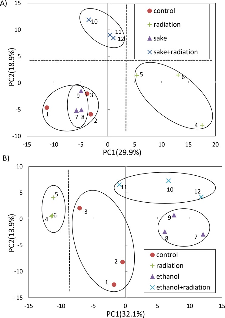
Metabolome analysis was next performed using CE-TOFMS to explore effects of sake on radiation-induced physiological alteration in the liver. In the analyses, a total of 230 (87 anions and 143 cations) and 245 metabolites (81 anions and 164 cations) were identified in the livers of mice administered sake and 15% ethanol, respectively (S3 and S4 Tables, respectively). PCA was performed to reveal differences in the metabolite profiles of the four treatment groups, which consisted of the controls, radiation, sake (or 15% ethanol) and the combination of both alcohol administration (sake or 15% ethanol) and radiation (Fig 2). In the PCA score plot, the group that received a combination of radiation and sake was clearly separated from the other three groups by the second principal component (PC2, 18.9% proportion; Fig 2A). The radiation and other groups appeared to be separated slightly along the first principal component (PC1, 29.9% proportion). The PCA score plot indicated that the metabolic profiles among individuals in each group were highly similar. A heat map representation of HCA (S3 Fig) also showed that clear differences were detectable among four groups and that individuals in each group have similar characteristics. In the case of 15% ethanol administration (Fig 2B), the group that received a combination of radiation and 15% ethanol was not clearly separated from the other groups along either PC1 or PC2. This lack of separation was in contrast to the group that received a combination of radiation and sake (Fig 2A). The PCA plot appears to show that 15% ethanol and radiation influence metabolic alterations of the liver in an independent manner, although the radiation and other three treatments groups were separated along PC1 (Fig 2B). Taken together, these results suggest that sake has distinct effects from ethanol on the influences of radiation on the liver metabolism.
Fig 2. PCA of metabolic data for the combined effects of sake (or ethanol) and radiation on livers.
(A) PCA in sake administration experiment, (B) PCA in 15% ethanol administration experiment. Percentage values indicated on the axes represent the contribution rate of the first (PC1) and second (PC2) principal components.
Effects of sake on the liver metabolome of irradiated mice
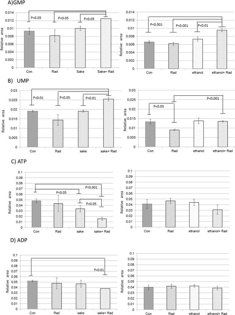
Using the correlation coefficients between the PC scores and variables for factor loading [22], we attempted to identify liver metabolites in irradiated mice were affected by sake administration. Metabolites that reached significant levels (p<0.01) in the evaluation of positive and negative correlation using the correlation coefficients were selected from the PC2 data generated from mice treated with a combination of radiation and sake (S5 Table). Among the selected metabolites, several were related to purine and pyrimidine metabolism and included AMP GMP, UMP, and CMP, which were found to be influenced by radiation. The relative area levels of GMP and UMP, which are representative nucleotide monophosphates, are shown in Fig 3A and 3B. Interestingly, although the diphosphates and triphosphates of adenosine (Fig 3C and 3D), guanosine, and uridine were not selected from PC2 data (S5 Table), the levels of these metabolites decreased in the livers of mice exposed to sake and radiation (S3 Table). Among the selected metabolites (S5 Table), seven metabolites (3-dephospho-CoA, GSH, nicotineamide, cysteine glutathione disulfide, GMP, UMP, and sedoheptulose 7-phosphate) were significantly modulated in the livers of mice treated with radiation and sake compared to the levels in the control, and sake and radiation alone-treated mice (S3 Table). In contrast, no changes in ATP, or ADP were detected in the livers of mice treated with the combination of radiation and ethanol (Fig 3C and 3D). Based on these findings for nucleotide metabolites, it appears that only guanosine nucleotide (GTP, GDP, and GMP) metabolism among nucleotides is influenced by radiation in the cases of both sake and ethanol. Although ATP depletion in combination of radiation and sake could result from hepatic failure induced by radiation or oxidative damages [23], taking it into consideration that a decrease in glutathione (GSH) is detected in liver diseases [24] but is not observed here as mentioned in the following part, it may be influenced by energy consumption for protection of livers or recovery from damages. In the livers of mice exposed to radiation and sake, the changes in several metabolites involved in glycolysis and pentose phosphate cycle, including glucose-6-phospate and sedoheptulose 7-phosphate, but not in the TCA cycle, were observed (S5 Table). ATP depletion may also be influenced by activities of these energy cycles. In addition, as nucleotide metabolism is known to be active in proliferating cells, including those of regenerating livers, the observed increases in several metabolites related to purine and pyrimidine metabolism such as AMP or GMP may be required for the repair and recovery of livers from damages [25–27]. Notably, as the increase in UMP was only observed in the liver of mice treated with a combination of sake and radiation, UMP might be involved in the metabolism of sugars present in sake [28].
Fig 3. Effects of sake or ethanol on radiation-induced alterations of purine and pyrimidine metabolism in livers.
(A) GMP, (B) UMP, (C) ATP, and (D) ADP. Data are relative area values of the metabolites and are presented as means ± S.D. of triplicate samples. Statistical analyses were performed by Welch’s t-test.
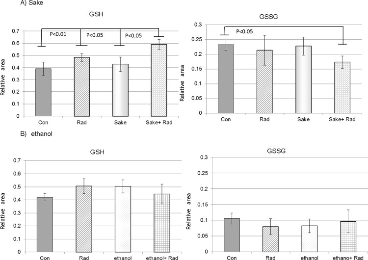
Among the seven selected metabolites that were significantly modulated in the livers of mice treated with radiation and sake, GSH is an important regulator of redox homeostasis and GSH/GSSG (glutathione disulfide) is considered to be the major redox couple that determines anti-oxidative capacity. GSSG is the oxidized form of GSH, and the GSH/GSSG ratio is often used as an indicator of the cellular redox state. Here, the levels of GSH and GSSG significantly increased and decreased, respectively, in the livers of mice treated with a combination of radiation and sake (Fig 4). The changes in these metabolites were not observed in mice administered 15% ethanol instead of sake (Fig 4), suggesting that glutathione metabolism is specifically influenced by the consumption of sake.
Fig 4. Effects of sake or ethanol on radiation-induced changes of GSH and GSSG in mouse livers.
(A) Sake and (B) ethanol. Data are relative area values of the metabolites and are presented as means ± S.D. of triplicate samples. Statistical analyses were performed by Welch’s t-test.
Characterization of radiation-induced metabolic alterations in livers
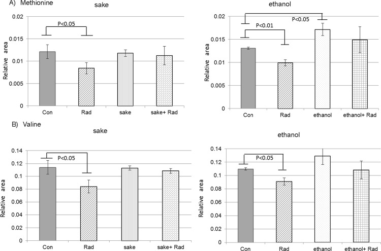
Metabolites that have significant correlation between the PC1 score and the relative peak area values were also selected in PC1, which characterized the radiation-alone group in the experiment examining sake and radiation (Fig 2A and S6 Table). The relative peak area values of 25 metabolites in the selected metabolites were significantly altered by radiation (S3 and S6 Tables). Among these metabolites, seven metabolites (3-methylhistidine, Val, Met, γ-butyrobetaine, N6-methyllysine, UDP-glucuronic acid, and UDP-glucose/UDP-galactose), which were also significantly changed on the relative peak area value levels in the livers of mice treated with radiation alone compared to the control in the experiment using 15% ethanol and radiation, were identified. These metabolites appeared to be altered stably and specifically in response to radiation in the livers of mice. With the exception of UDP-glucose/UDP-galactose, all of the metabolites decreased in response to radiation exposure alone. Radiation induced a decrease in the levels of methionine (Fig 5A), a finding that was previously reported [29], suggesting that alteration of methionine metabolism may be related to carcinogenesis [30,31]. The radiation-induced decrease in methionine levels was restored to control levels by the administration of sake or 15% ethanol. Although the administration of sake alone had no effect on the methionine level, sake administration diminished the effect of radiation on the levels of methionine. In the case of 15% ethanol, methionine levels induced by ethanol may have influenced the results (Fig 5A). It was reported that alcohol consumption impairs various methylation reactions in the liver [32]. However, a decrease in methionine in the livers of mice administered sake or ethanol alone was not observed. In contrast, the decrease in methionine induced by radiation was suppressed to control levels in irradiated mice after treatment with sake or ethanol. The contrasting results from these studies may be related to differences between mouse strains, sexes, or diets, with respect to the regulation of methylation reactions related to metabolism [33,34]. Further studies are therefore needed to determine the mechanisms underlying the mediation of methylation metabolism by sake or ethanol. Radiation was also shown to induce a decrease in the valine content of livers, which appeared to be influenced by the administration of sake or 15% ethanol (Fig 5B). Valine depletion is suggested to be associated with mTOR /S6K signaling suppression [35]. Interestingly, mTOR is related to reactive oxygen species (ROS) signaling [36]. Therefore, ROS induced by radiation might induce valine depletion, which in turn leads to the suppression of mTOR signaling. In addition, as supplementation of branched-chain amino acids, including valine, appears to reduce radiation-induced damage [37], the suppression of the valine depletion by exposure to sake or ethanol suggest that alcohol administration has protective abilities.
Fig 5. Changes in methionine and valine in the livers of irradiated mice administered sake or ethanol.
(A) Methionine and (B) valine. Data are relative area values of the metabolites, and are presented as means ± S.D. of triplicate samples. Statistical analyses were performed using Welch’s t-test.
Here, metabolites that exhibited significant (P<0.01) trends using correlation coefficients between the PC scores and variables for factor loading in PC2 or PC1 as indicated in S5 and S6 Tables were compared. In addition, although the ethanol and sake administration were performed independently, metabolite levels in the control and radiation groups were measured in both experiments. As relative values were obtained in each experiment, they cannot be combined for the statistical analyses; however, similar changes in metabolite levels were detected in the control and radiation groups between two independent experiments. These results indicate that this experimental approach provides consistent results and allows changes in the selected metabolites to be evaluated.
Alteration of metabolic biochemical markers in serum
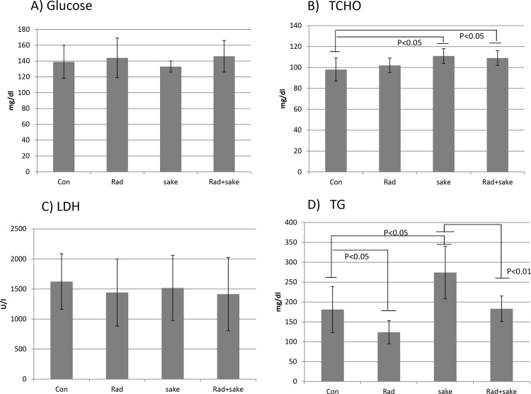
Changes in the serum levels of several metabolic biochemical markers that accompanied by alterations in liver metabolism in the four treatment groups were next evaluated. The serum levels of lactose dehydrogenase (LDH) and glucose were not markedly changed in mice treated with sake and/or radiation compared to the control group. Although the serum level of total cholesterol (TCHO) increased in mice administered sake alone, radiation had no effect on the level of TCHO; however, that of triglycerides (TG) increased in mice administered sake alone, and was reduced to control levels following irradiation (Fig 6). In this experiment, the amount of sake administered to mice seemed to be excessive because a significant increase in serum TG was observed compared to control mice. Although radiation alone induced a reduction in TG levels, the serum TG level in the treatment group that received both sake and radiation is greatly reduced to the control level from the level in mice administered sake. The observed reduction of TG by radiation in mice administered sake may be in part due to an induction of anti-oxidative responses, as indicated by the increase in GSH in the liver. The alcohol-induced accumulation of TG can reportedly be mitigated by a diet including foods that contain factors that promote anti-oxidative responses [38].
Fig 6. Effects of sake on metabolic biochemical markers in the serum of irradiated mice.
(A) Glucose, (B) total cholesterol (TCHO), (C) lactose dehydrogenase (LDH), and (D) triglycerides (TG). Data are presented as means ± S.D. from seven mice in two independent experiments. Statistical analyses were performed by the unpaired t-test.
Influence of sake administration on the effects of radiation
It has been reported that wine consumption mitigates the side effects associated with radio-cancer therapies [39] and that beer consumption can reduce the adverse effects of radiation [40]. However, a greater understanding of the effects of alcoholic beverages on responses to radiation is needed for assessing radiation risk or medical applications of alcohol. An omics-based approach was used to examine molecular changes in the livers of rats treated with sake [41]. However, such approaches have not been applied to the determination of the specific effects of sake on liver metabolism versus those of ethanol. To the best of our knowledge, metabolic analyses have not been performed from the viewpoint of mediation of a stress to other stress effects. Sake has anti-mutagenic effects, a property that has not been attributed to ethanol [14], and the administration of acute doses of sake has been shown to protect mice from the adverse effects of high-dose radiation more effectively than ethanol alone [15].
Sake, which is brewed from rice, water, and rice koji mold, contains numerous components, including sugars, amino acids, and vitamins [42]. GSH is induced by various components in foods, including rice proteins, and the effects we found seem to be due to the involved amino acid components in sake [43]. The induction of anti-oxidative activity is considered to be influenced by amino acids, including cysteine. The free amino acid composition of the sake used in this study was analyzed and was found to be comprised of relatively high levels of alanine, glutamate, and glycine, which are related to GSH synthesis (Table 1) [44]. Recently, it was demonstrated that glutamine and alanine induce anti-oxidative activity in livers [45]. In the present study, although the administration of sake alone had no marked effects on GSH regulation, and glutamine was not abundantly present in the sake (Table 1), the amino acid components of sake may be related to the altered regulation of GSH that was observed in irradiated mice after sake administration. The sake used in the present study and that evaluated in a report that demonstrated sake mitigates high-dose radiation effects [15] shared common characteristics, such as the abundance of alanine, glutamate, and glycine. Thus, it is possible that these amino acids are partly responsible for the beneficial effects of sake on radiation-induced damages.
Table 1. Free amino acids in sake (mg per 100g).
| Amino acids | Amounts (mg) per 100g sake | Amino acids | Amounts (mg) per 100g sake |
|---|---|---|---|
| Asp | 3 | Cys | 1 |
| Thr | 2 | Met | <1 |
| Ser | 3 | Ile | 2 |
| Asn | 4 | Leu | 5 |
| Glu | 9 | Tyr | 5 |
| Gln | <1 | Phe | 2 |
| Pro | 10(±1.2) | His | 2 |
| Gly | 8 | Lys | 2 |
| Ala | 13(±1.2) | Trp | <1 |
| Val | 4 | Arg | 3 |
Data are presented as means (mg) of three samples from three different lots. Means (±SD) in the case of SD>1.
Although irradiation and sake administration were clearly shown to have interactive effects with respect to GSH regulation, the underlying mechanism remains unclear. We previously demonstrated that obesity mediates radiation sensitivity [8]. The harmful effects of radiation on living organisms, particularly in the case of low-linear energy transfer (LET) radiation, such as X-rays, are considered to result from radiation-induced oxidative stress. In the case of obesity, obesity-induced oxidative stress seems to mediate the effects of radiation in livers. Sake may influence redox homeostasis in the liver by a yet-unidentified mechanism, resulting in the alteration of GSH regulation following exposure to radiation.
Although the biological effects of alcohol administration have been investigated in various experimental models [32, 38, 46, 47], in the present study, we exposed mice to alcohol for one month as an experimental model. Chronic alcohol administration for 2 or 6 weeks has been used to evaluate the effects of alcohol on metabolism in mice [32, 38, 47], and consistent with these models, an increase in TG was also observed here, indicating that this model is considered as an appropriate model to evaluate chronic alcohol administration.
For the evaluation of factors in mechanisms related to liver diseases, it is necessary to identify the proteins and metabolites that are altered under various conditions [48]. Metabolome analysis provides insight into the underlying mechanisms that lead to the development of diseases, such as cancers, and may lead to the identification of potential therapeutic targets [49]. In the case of alcohol-related disease, a relationship between chronic alcohol consumption and fatty liver has been confirmed [2]. In addition, the development of fatty liver appears to be influenced by radiation [50]. The present findings that sake consumption mitigates the effects of radiation may provide insight into relationship between alcohol-related diseases and radiation effects.
As an experimental model, we used mice administered either sake or ethanol that were then exposed to fractionated irradiation (0.75 Gy, 4 times). Fractionated irradiation with approximately 0.75 Gy has been performed at various intervals in several studies [51–53]. The dose per fraction used here was similar to the lowest value used in a report evaluating the combined effects of radiation and chemical treatment on carcinogenesis [51]. Compared to single irradiation, fractionated irradiation increases the potential that radiation-related effects that are altered in response to chronic sake administration can be detected. In the present model, to evaluate effects of radiation on mice exposed to chronic alcohol intake, the fractionated irradiation was performed during the last week of the intake period. In addition, because the fractionated dose has been used in experimental models for evaluating the effects of radiation on normal cells or carcinomas in radiation therapies [52, 53], the results from the present study may contribute to medical applications. Though the development of second primary cancers after radiation therapy may be influenced by lifestyles, there are limited data for the influence of specific factors, with the exception of tobacco, on their epidemiology [54]. For this reason, the National Council on Radiation Protection & Measurements (NCRP) has recommended that the relationships between second primary cancers and lifestyles should be investigated [55,56].
Alcohol consumption represents a type of caloric intake and considered to influence body weight, although this relationship is controversial [57, 58]. Although the final body weights of the mice administered ethanol or sake did not significantly differ from that of the control group (S1 and S2 Tables), fluctuations in body weights were observed during the administration period. In particular, clear decreases in mice administered 15% ethanol or sake were detected approximately two to three weeks after the start of the administration period. As decreases in food intake in mice administered 15% ethanol were observed during this period (S7 Table), the observed decreases in body weight were likely related to decreased food intake. Although the reduction in food intake appeared to have recovered at the end of the administration period, the observed changes in metabolites or TG may have resulted from alterations in calorie consumption after alcohol administration.
Drinking sake in moderation has beneficial and protective health effects, and may provide partial protection against accidental or medical radiation exposure. Although the amount of sake administration in this study is not small, the level appears to induce protective effects and is practical for evaluation in mouse experimental model [15]. If the natural components in sake that promote these protective effects, are identified, they may contribute to the understanding of the benefits of certain foods on health and potentially be used in the clinical setting. To this end, evaluation of effects of single irradiation on metabolisms or identification of metabolic markers by lipidomics would be needed. The findings presented here warrant the further study of the beneficial components present in sake and analysis of the metabolic networks altered by sake consumption.
Conclusions
Chronic Japanese sake consumption induces specific metabolic alterations in the liver in response to irradiation. Although excess sake consumption may induce adverse effects on the liver, sake intake has the potential to promote anti-oxidative stress activities following radiation exposure. The findings presented here suggest that moderate sake consumption may promote anti-oxidative activity following exposure to stress such as radiation, thereby limiting the adverse effects typically associated with these stresses.
Supporting Information
(PDF)
(PDF)
The X-axis is labeled with group names and sample numbers and the Y-axis shows peaks. Sample numbers on the X-axis correspond to the numbers in Fig 2A. Peaks were analyzed by HCA and distances are depicted by a tree diagram. In the color legend, the lowest value in the map is represented by a bright green, and the highest value is represented by a bright red.
(PDF)
(PDF)
(PDF)
Values represent the relative areas of each metabolite peak to the peak area of an internal standard.
(PDF)
Values represent the relative areas of each metabolite peak to the peak area of an internal standard.
(PDF)
These have significant correlation (P<0.01) based on factor loadings between the PC2 scores and variables in the metabolome data for irradiated mice administered sake.
(PDF)
These have significant correlation (P<0.01) based on factor loadings between the PC1 scores and variables in the metabolome data for irradiated mice administered sake.
(PDF)
(PDF)
Acknowledgments
The authors would like to thank Ms. Kyoko Sakuma, Ms. Taeko Iwai, Ms. Hiromi Arai and Ms. Yasuko Morimoto for their expert technical assistance and administrative support.
Data Availability
All relevant data are within the paper and its Supporting Information files.
Funding Statement
The authors received no specific funding for this work. The work was performed by the author's institution's budget.
References
- 1.Khan N, Afaq F, Mukhtar H. Lifestyle as risk factor for cancer: Evidence from human studies. Cancer Lett. 2010; 293: 133–143. 10.1016/j.canlet.2009.12.013 [DOI] [PMC free article] [PubMed] [Google Scholar]
- 2.Orman ES, Odena G, Bataller R. Alcoholic liver disease: Pathogenesis, management, and novel targets for therapy. J Gastroenterol Hepatol. 2013; 28: 77–84. 10.1111/jgh.12030 [DOI] [PMC free article] [PubMed] [Google Scholar]
- 3.Donato F, Tagger A, Gelatti U, Parrinello G, Boffetta P, Albertini A, et al. Alcohol and hepatocellular carcinoma: the effect of lifetime intake and hepatitis virus infections in men and women. Am J Epidemiol. 2002; 155. [DOI] [PubMed] [Google Scholar]
- 4.Schottenfeld D. Alcohol as a cofactor in the etiology of cancer. Cancer. 1979; 43: 1962–1966. [DOI] [PubMed] [Google Scholar]
- 5.Poschl G, Seitz HK. Alcohol and cancer. Alcohol Alcohol. 2004; 39: 155–165. [DOI] [PubMed] [Google Scholar]
- 6.Marrero JA, Fontana RJ, Fu S, Conjeevaram HS, Su GL, Lok AS. Alcohol, tobacco and obesity are synergistic risk factors for hepatocellular carcinoma. J Hepatol. 2005; 42: 218–224. [DOI] [PubMed] [Google Scholar]
- 7.Inoue M, Tsugane S, JPHC Study Group. Impact of alcohol drinking on total cancer risk: data from a large-scale population-based cohort study in Japan. Br J Cancer. 2005; 92: 182–187. [DOI] [PMC free article] [PubMed] [Google Scholar]
- 8.Vares G, Wang B, Ishii-Ohba H, Nenoi M, Nakajima T. Diet-induced obesity modulates epigenetic responses to ionizing radiation in mice. PLoS One. 2014; 9: e106277 10.1371/journal.pone.0106277 [DOI] [PMC free article] [PubMed] [Google Scholar]
- 9.Yoshida K, Inoue T, Nojima K, Hirabayashi Y, Sado T. Calorie restriction reduces the incidence of myeloid leukemia induced by a single whole-body radiation in C3H/He mice. Proc Natl Acad Sci U S A. 1997; 94: 2615–2619. [DOI] [PMC free article] [PubMed] [Google Scholar]
- 10.Shang Y, Kakinuma S, Yamauchi K, Morioka T, Kokubo T, Tani S, et al. Cancer prevention by adult-onset calorie restriction after infant exposure to ionizing radiation in B6C3F1 male mice. Int J Cancer. 2014; 135: 1038–1047. 10.1002/ijc.28751 [DOI] [PubMed] [Google Scholar]
- 11.Iwasaki T, Murata M, Ohshima S, Miyake T, Kudo S, Inoue Y, et al. Second analysis of mortality of nuclear industry workers in Japan,1986–1997. Radiat Res. 2003; 159: 228–238. [DOI] [PubMed] [Google Scholar]
- 12.Akiba S, Mizuno S. The third analysis of cancer mortality among Japanese nuclear workers, 1991–2002: estimation of excess relative risk per radiation dose. J Radiol Prot. 2012; 32: 73–83. 10.1088/0952-4746/32/1/73 [DOI] [PubMed] [Google Scholar]
- 13.Wynder EL, Hoffmann D, McCoy GD, Cohen LA, Reddy BS. Tumor promotion and cocarcinogenesis as related to man and his environment In: Slaga TJ, Sivak A, Boutwell RK, editors. Carcinogenesis: Mechanisms of tumor promotion and cocarcinogenesis, vol2 New York: Raven Press; 1978. pp. 59–77. [Google Scholar]
- 14.Arimoto-Kobayashi S, Sugiyama C, Harada N, Takeuchi M, Takemura M, Hayatsu H. Inhibitory effects of beer and other alcoholic beverages on mutagenesis and DNA adduct formation induced by several carcinogens. J Agric Food Chem. 1999; 47: 221–230. [DOI] [PubMed] [Google Scholar]
- 15.Takizawa Y, Yamashita J, Ishigouka S. Protective effect of Japanese sake against ionizing X-irradiation in mice. Radioisotopes. 2014; 63: 1–12. [Google Scholar]
- 16.Gu X, Manautou JE. Molecular mechanisms underlying chemical liver injury. Expert Rev Mol Med. 2012; 14: e4 10.1017/S1462399411002110 [DOI] [PMC free article] [PubMed] [Google Scholar]
- 17.Nakajima T. Roles of sulfur metabolism and rhodanese in detoxification and anti-oxidative stress functions in the liver: responses to radiation exposure. Med Sci Monit. 2015; 21: 1721–1725. 10.12659/MSM.893234 [DOI] [PMC free article] [PubMed] [Google Scholar]
- 18.Lieber CS. Relationships between nutrition, alcohol use, and liver disease. Alcohol Res Health. 2003; 27: 220–231. [PMC free article] [PubMed] [Google Scholar]
- 19.Kim SR, Kubo T, Kuroda Y, Hojyo M, Matsuo T, Miyajima A, et al. Comparative metabolome analysis of cultured fetal and adult hepatocytes in humans. J Toxicol Sci. 2014; 39: 717–723. [DOI] [PubMed] [Google Scholar]
- 20.Takahashi S, Saito K, Jia H, Kato H. An integrated multi-omics study revealed metabolic alterations underlying the effects of coffee consumption. PLoS One. 2014; 9: e91134 10.1371/journal.pone.0091134 [DOI] [PMC free article] [PubMed] [Google Scholar]
- 21.Goto T, Kubota Y, Toyoda A. Plasma and liver metabolic profiles in mice subjected to subchronic and mild social defeat stress. J Proteome Res. 2015; 14: 1025–1032. 10.1021/pr501044k [DOI] [PubMed] [Google Scholar]
- 22.Yamamoto H, Fujimori T, Sato H, Ishikawa G, Kami K, Ohashi Y. Statistical hypothesis testing of factor loading in principal component analysis and its application to metabolite set enrichment analysis. Bmc Bioinformatics. 2014; 15. [DOI] [PMC free article] [PubMed] [Google Scholar]
- 23.Nishikawa T, Bellance N, Damm A, Bing H, Zhu Z, Handa K, et al. A switch in the source of ATP production and a loss in capacity to perform glycolysis are hallmarks of hepatocyte failure in advance liver disease. J Hepatol. 2014; 60: 1203–1211. 10.1016/j.jhep.2014.02.014 [DOI] [PMC free article] [PubMed] [Google Scholar]
- 24.Czeczot H, Ścibior D, Skrzycki M, Podsiad M. Glutathione and GSH-dependent enzymes in patients with liver cirrhosis and hepatocellular carcinoma. Acta Biochim Pol. 2006; 53: 237–241. [PubMed] [Google Scholar]
- 25.Weber G, Prajda N, Jackson R. Key enzymes of IMP metabolism: transformation and proliferation-linked alterations in gene expression. Adv Enzyme Regul. 1976; 14: 3–24. [DOI] [PubMed] [Google Scholar]
- 26.Marijnen Y, de Korte D, Roos D, van Gennip A. Purine and pyrimidine metabolism of normal and leukemic lymphocytes. Adv Exp Med Biol. 1989; 253A: 433–438. [DOI] [PubMed] [Google Scholar]
- 27.Pausch J, Keppler D, Decker K. Activity and distribution of the enzymes of uridylate synthesis from orotate in animal tissues. Biochim Biophys Acta. 1972; 258: 395–403. [DOI] [PubMed] [Google Scholar]
- 28.Ishibashi Y, Hirabayashi Y. AMP-activated protein kinase suppresses biosynthesis of glucosylceramide by reducing intracellular sugar nucleotides. J Biol Chem. 2015. [DOI] [PMC free article] [PubMed] [Google Scholar]
- 29.Batra V, Verma P. Dietary L-methionine supplementation mitigates gamma-radiation induced global DNA hypomethylation: Enhanced metabolic flux towards S-adenosyl-L-methionine (SAM) biosynthesis increases genomic methylation potential. Food Chem Toxicol. 2014; 69: 46–54. 10.1016/j.fct.2014.03.040 [DOI] [PubMed] [Google Scholar]
- 30.Ghoshal A, Farber E. The induction of liver cancer by dietary deficiency of choline and methionine without added carcinogens. Carcinogenesis. 1984; 5: 1367–1370. [DOI] [PubMed] [Google Scholar]
- 31.Avila M, Berasain C, Torres L, Martín-Duce A, Corrales F, Yang H, et al. Reduced mRNA abundance of the main enzymes involved in methionine metabolism in human liver cirrhosis and hepatocellular carcinoma. J Hepatol. 2000; 33: 907–914. [DOI] [PubMed] [Google Scholar]
- 32.Kharbanda KK, Todero SL, Thomes PG, Orlicky DJ, Osna NA, French SW, et al. Increased methylation demand exacerbates ethanol-induced liver injury. Exp Mol Pathol. 2014; 97: 49–56. 10.1016/j.yexmp.2014.05.006 [DOI] [PubMed] [Google Scholar]
- 33.Christensen KE, Wu Q, Wang X, Deng L, Caudill MA, Rozen R. Steatosis in mice is associated with gender, folate intake, and expression of genes of one-carbon metabolism. J Nutr. 2010; 140: 1736–1741. 10.3945/jn.110.124917 [DOI] [PubMed] [Google Scholar]
- 34.Scott MC, Guerciolini R, Szumlanski C, Weinshilboum RM. Mouse kidney histamine N-methyltransferase: assay conditions, biochemical properties and strain variation. Agents Actions. 1991; 32: 194–202. [DOI] [PubMed] [Google Scholar]
- 35.Kakazu E, Kanno N, Ueno Y, Shimosegawa T. Extracellular branched-chain amino acids, especially valine, regulate maturation and function of monocyte-derived dendritic cells. J Immunol. 2007; 179: 7137–7146. [DOI] [PubMed] [Google Scholar]
- 36.Alexander A, Cai SL, Kim J, Nanez A, Sahin M, MacLean KH, et al. ATM signals to TSC2 in the cytoplasm to regulate mTORC1 in response to ROS. Proc Natl Acad Sci U S A. 2010; 107: 4153–4158. 10.1073/pnas.0913860107 [DOI] [PMC free article] [PubMed] [Google Scholar]
- 37.Lee IJ, Seong J, Bae JI, You SH, Rhee Y, Lee JH. Effect of oral supplementation with branched-chain amino acid (BCAA) during radiotherapy in patients with hepatocellular carcinoma: a double-blind randomized study. Cancer Res Treat. 2011; 43: 24–31. 10.4143/crt.2011.43.1.24 [DOI] [PMC free article] [PubMed] [Google Scholar]
- 38.Xiang J, Zhu W, Li Z, Ling S. Effect of juice and fermented vinegar from Hovenia dulcis peduncles on chronically alcohol-induced liver damage in mice. Food Funct. 2012; 3: 628–634. 10.1039/c2fo10266h [DOI] [PubMed] [Google Scholar]
- 39.Morganti AG, Digesu C, Panunzi S, De Gaetano A, Macchia G, Deodato F, et al. Radioprotective effect of moderate wine consumption in patients with breast carcinoma. Int J Radiat Oncol Biol Phys. 2009; 74: 1501–1505. 10.1016/j.ijrobp.2008.10.089 [DOI] [PubMed] [Google Scholar]
- 40.Monobe M, Ando K. Drinking beer reduces radiation-induced chromosome aberrations in human lymphocytes. J Radiat Res. 2002; 43: 237–245. [DOI] [PubMed] [Google Scholar]
- 41.Masuo Y, Imai T, Shibato J, Hirano M, Jones OAH, Maguire ML, et al. Omic analyses unravels global molecular changes in the brain and liver of a rat model for chronic Sake (Japanese alcoholic beverage) intake. Electrophoresis. 2009; 30: 1259–1275. 10.1002/elps.200900045 [DOI] [PubMed] [Google Scholar]
- 42.Mimura N, Isogai A, Iwashita K, Bamba T, Fukusaki E. Gas chromatography/mass spectrometry based component profiling and quality prediction for Japanese sake. J Biosci Bioeng. 2014; 118: 406–414. 10.1016/j.jbiosc.2014.04.006 [DOI] [PubMed] [Google Scholar]
- 43.Yang L, Chen JH, Xu T, Zhou AS, Yang HK. Rice protein improves oxidative stress by regulating glutathione metabolism and attenuating oxidative damage to lipids and proteins in rats. Life Sci. 2012; 91: 389–394. 10.1016/j.lfs.2012.08.003 [DOI] [PubMed] [Google Scholar]
- 44.Wu G, Fang YZ, Yang S, Lupton JR, Turner ND. Glutathione Metabolism and Its Implications for Health. J Nutr 2004; 134: 489–492. [DOI] [PubMed] [Google Scholar]
- 45.Cruzat VF, Bittencourt A, Scomazzon SP, Moreira Leite JS, Homem de Bittencourt PI Jr., Tirapegui J. Oral free and dipeptide forms of glutamine supplementation attenuate oxidative stress and inflammation induced by endotoxemia. Nutrition. 2014; 30: 602–611. 10.1016/j.nut.2013.10.019 [DOI] [PubMed] [Google Scholar]
- 46.Phillips BJ, Jenkinson P. Is ethanol genotoxic? A review of the published data. Mutagenesis. 2001; 16: 91–101. [DOI] [PubMed] [Google Scholar]
- 47.Lee HI, Seo KO, Yun KW, Kim MJ, Lee MK. Comparative study of the hepatoprotective efficacy of Artemisia iwayomogi and Artemisia capillaris on ethanol-administered mice. J Food Sci. 2011; 76: T207–211. 10.1111/j.1750-3841.2011.02385.x [DOI] [PubMed] [Google Scholar]
- 48.Deaciuc IV, Song Z, McClain CJ. Lessons from large-scale gene profiling of the liver in alcoholic liver disease. Hepatol Res. 2005; 31: 187–192. [DOI] [PubMed] [Google Scholar]
- 49.Puszyk WM, Trinh TL, Chapple SJ, Liu C. Linking metabolism and epigenetic regulation in development of hepatocellular carcinoma. Lab Invest. 2013; 93: 983–990. 10.1038/labinvest.2013.94 [DOI] [PMC free article] [PubMed] [Google Scholar]
- 50.Akahoshi M, Amasaki Y, Soda M, Hida A, Imaizumi M, Nakashima E, et al. Effects of radiation on fatty liver and metabolic coronary risk factors among atomic bomb survivors in Nagasaki. Hypertens Res. 2003; 26: 965–970. [DOI] [PubMed] [Google Scholar]
- 51.Kakinuma S, Nishimura M, Amasaki Y, Takada M, Yamauchi K, Sudo S, et al. Combined exposure to X-irradiation followed by N-ethyl-N-nitrosourea treatment alters the frequency and spectrum of Ikaros point mutations in murine T-cell lymphoma. Mutat Res. 2012; 737: 43–50. 10.1016/j.mrfmmm.2012.06.001 [DOI] [PubMed] [Google Scholar]
- 52.Joiner MC, Denekamp J, Maughan RL. The use of ‘top-up’ experiments to investigate the effect of very small doses per fraction in mouse skin. Int J Radiat Biol Relat Stud Phys Chem Med. 1986; 49: 565–580. [DOI] [PubMed] [Google Scholar]
- 53.Wrenn DC, Saigai K, Lucci JA 3rd, Pearson MJ, Simpkins F, Schuman S, et al. A phase I study using low-dose fractionated whole abdominal radiotherapy as a chemopotentiator to full-dose cisplatin for optimally debulked stage III/IV carcinoma of the endometrium. Gynecol Oncol. 2011; 122: 59–62. 10.1016/j.ygyno.2011.03.007 [DOI] [PubMed] [Google Scholar]
- 54.Wood ME, Vogel V, Ng A, Foxhall L, Goodwin P, Travis LB. Second malignant neoplasms: assessment and strategies for risk reduction. J Clin Oncol. 2012; 30: 3734–3745. 10.1200/JCO.2012.41.8681 [DOI] [PubMed] [Google Scholar]
- 55.Travis LB, Ng AK, Allan JM, Pui CH, Kennedy AR, Xu XG, et al. Second malignant neoplasms and cardiovascular disease following radiotherapy. J Natl Cancer Inst. 2012; 104: 357–370. 10.1093/jnci/djr533 [DOI] [PMC free article] [PubMed] [Google Scholar]
- 56.NCRP. (National council on radiation protection and measurements) Scientific Committee 1–17. Second Primary Cancers and Cardiovascular Disease After Radiotherapy. NCRP Report. 2011; No. 170.
- 57.Sayon-Orea C, Martinez-Gonzalez MA, Bes-Rastrollo M. Alcohol consumption and body weight: a systematic review. Nutrition Reviews. 2011; 69: 419–431. 10.1111/j.1753-4887.2011.00403.x [DOI] [PubMed] [Google Scholar]
- 58.Aguiar AS, Da-Silva VA, Boaventura GT. Can calories from ethanol contribute to body weight preservation by malnourished rats. Braz J Med Biol Res. 2004; 37: 841–846. [DOI] [PubMed] [Google Scholar]
Associated Data
This section collects any data citations, data availability statements, or supplementary materials included in this article.
Supplementary Materials
(PDF)
(PDF)
The X-axis is labeled with group names and sample numbers and the Y-axis shows peaks. Sample numbers on the X-axis correspond to the numbers in Fig 2A. Peaks were analyzed by HCA and distances are depicted by a tree diagram. In the color legend, the lowest value in the map is represented by a bright green, and the highest value is represented by a bright red.
(PDF)
(PDF)
(PDF)
Values represent the relative areas of each metabolite peak to the peak area of an internal standard.
(PDF)
Values represent the relative areas of each metabolite peak to the peak area of an internal standard.
(PDF)
These have significant correlation (P<0.01) based on factor loadings between the PC2 scores and variables in the metabolome data for irradiated mice administered sake.
(PDF)
These have significant correlation (P<0.01) based on factor loadings between the PC1 scores and variables in the metabolome data for irradiated mice administered sake.
(PDF)
(PDF)
Data Availability Statement
All relevant data are within the paper and its Supporting Information files.


