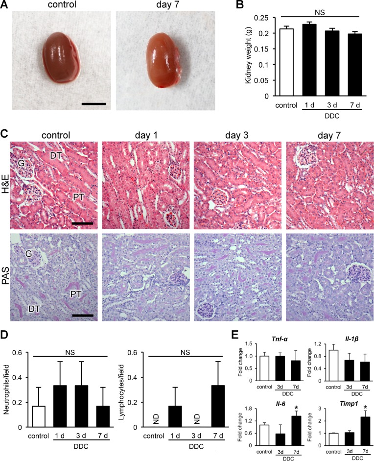
Fig 2. Kidneys of 3,5-diethoxycarbonyl-1,4-dihydrocollidine (DDC)-fed mice do not show definitive morphological changes on routine histological examination.
(A) Representative macroscopic photographs of the kidneys. Scale bar = 5 mm. (B) Kidney weights in control (white bar) and DDC-fed mice (black bar) at days 1, 3, and 7. (C) Histopathological images of kidneys of control and DDC-fed mice at days 1, 3, and 7 (top row, hematoxylin and eosin [H&E] staining; bottom row, periodic acid-Schiff [PAS] staining). PT, proximal tubules; DT, distal tubules; G, glomeruli. Scale bars = 100 μm. (D) Counts of Ly-6G/-6C-positive neutrophils and leukocyte common antigen (LCA)–positive lymphocytes in control (white bar) and DDC-fed mice (black bar) at days 1, 3, and 7. Data are presented as means ± SEM. NS, not significant; ND, not detected. (E) Quantitative reverse transcription polymerase chain reaction analyses for tumor necrosis factor-α (Tnf-α), interleukin-1β (Il-1β), interleukin-6 (Il-6), and tissue inhibitor of metalloproteinase 1 (Timp1) mRNA in control and DDC-fed mice at days 3 and 7. Values are normalized to Gapdh expression. The mRNA values were analyzed by the two-tailed Student’s t test. Data are presented as means ± SD. *P < 0.05 compared with control mice.

