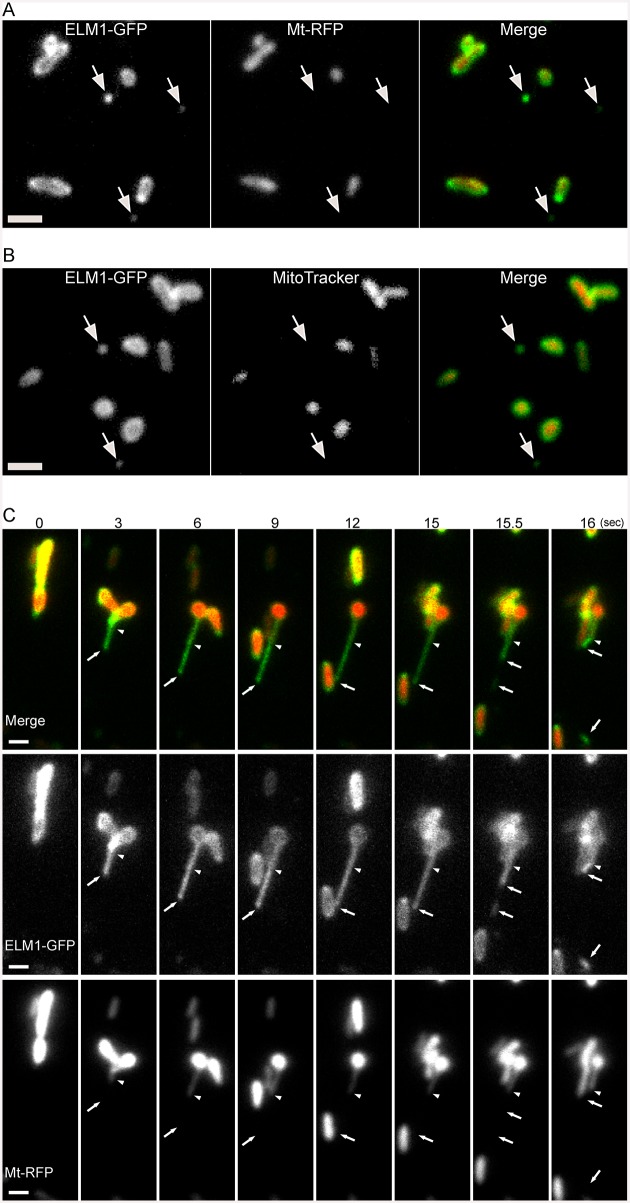
Fig 1. Microscopic observations of mitochondrial outer membrane, MDVs and MOPs.
(A) Images of Arabidopsis leaf epidermal cells expressing both ELM1-GFP and Mt-RFP. Fluorescent fusions of ELM1 were expressed under the control of the ELM1 promoter. The arrows indicate MDVs. Bar = 1 μm. (B) Images of Arabidopsis leaf epidermal cells transfected with ELM1-GFP and stained by using a mitochondrial inner membrane marker, MitoTracker. Bar = 1 μm. The arrows indicate MDVs. (C) Time-course observation of the formation of a MDV. These are representative images from a movie (S1 Movie) that was recorded at 10 frames per second. The arrows indicate a MDV and the tip of a MOP, and the arrowheads indicate the tip of a protruding matrixule. The time stamp is in seconds. Bars = 1 μm.

