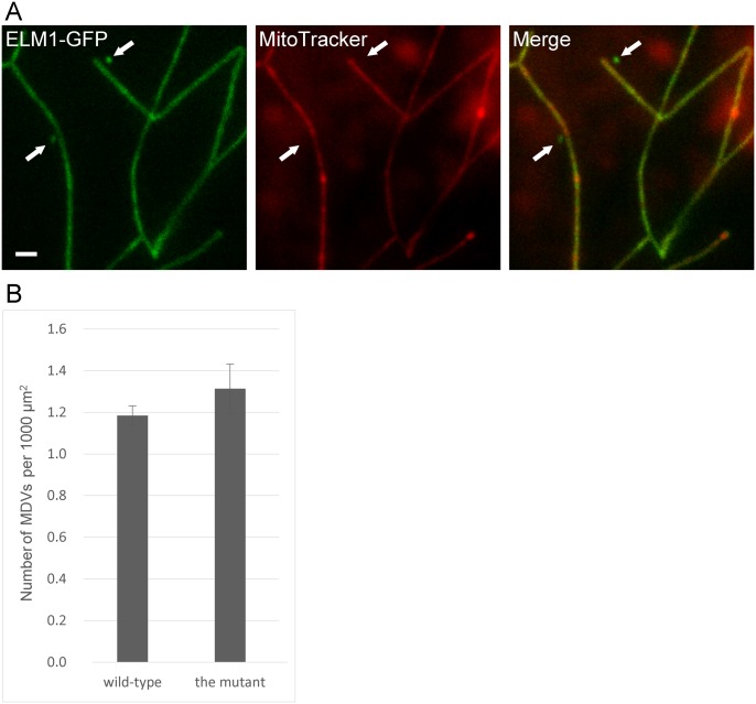
Fig 3. Microscopic observation of MDVs in the mitochondrial fission-defective mutant, drp3a drp3b.
(A) The T-DNA insertion mutant, drp3a drp3b, transfected with ELM1-GFP was stained with MitoTracker. The arrows indicate MDVs. Bar = 1 μm. (B) The numbers of MDVs in the double mutant and in wild-type. Images of 20 epidermal cells were analyzed with three replicates. Error bars represent standard error (SE).

