
Figure 2.
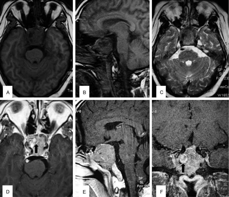
MRI of the brain revealed a large pituitary tumor that extended superiorly in the suprasellar cistern to elevate the optic chiasm and extended inferiorly to fill the sphenoid sinus. (A, B) The mass was isointense to brain parenchyma on T1-weighted MRI, (C) relatively hypointense on T2-weighted MRI. (D-F) Showed significant contrast enhancement after injection of gadolinium. There was abnormal signal intensity consistent with acute hemorrhage in the tumor (black arrow).