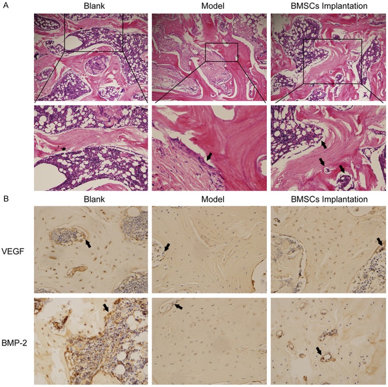
Figure 4.

BMSCs implanted into the ONFH rabbits. BMSCs implanted into the ONFH rabbit for 4 weeks. The femoral head fragments were collected and sectioned. A. The femoral head tissues were analyzed by H&E staining. Black arrows depicted OB and capillary. B. VEGF and BMP-2 expressions were examined by immunohistochemistry.
