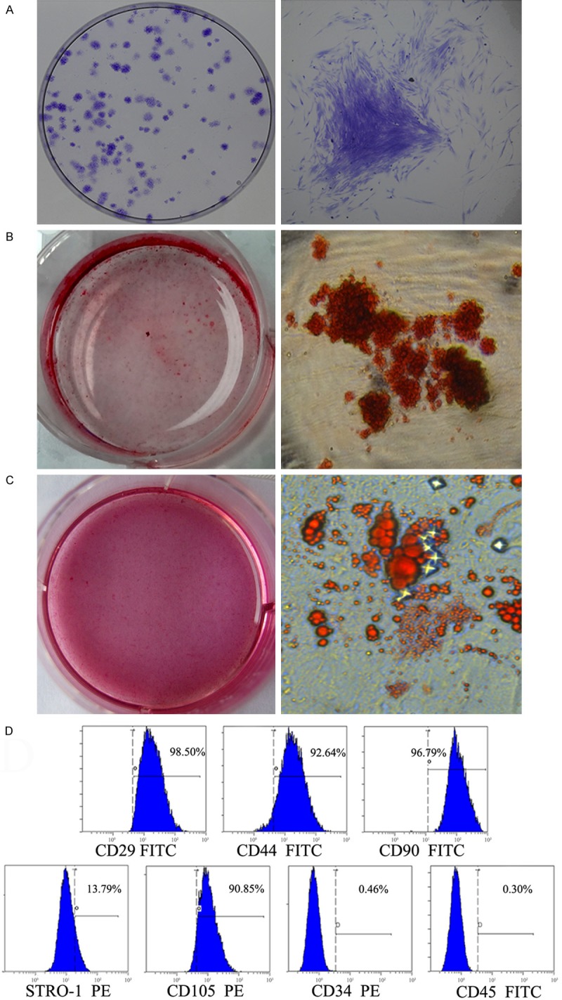
Figure 1.

Identification and characters of PDLSCs. (A) Representative images of colony-forming units from PDLSCs at 14 days. (B, C) Multipotent differentiation of PDLSCs. Osteogenic differentiation of PDLSCs was demonstrated by the presence of alizarin red S-positive mineralized nodules (B). Adipogenic differentiation PDLSCs was demonstrated by the formation of oil red O-positive lipid globules (C). (D) Flow-cytometric analysis of surface markers expressed on PDLSCs.
