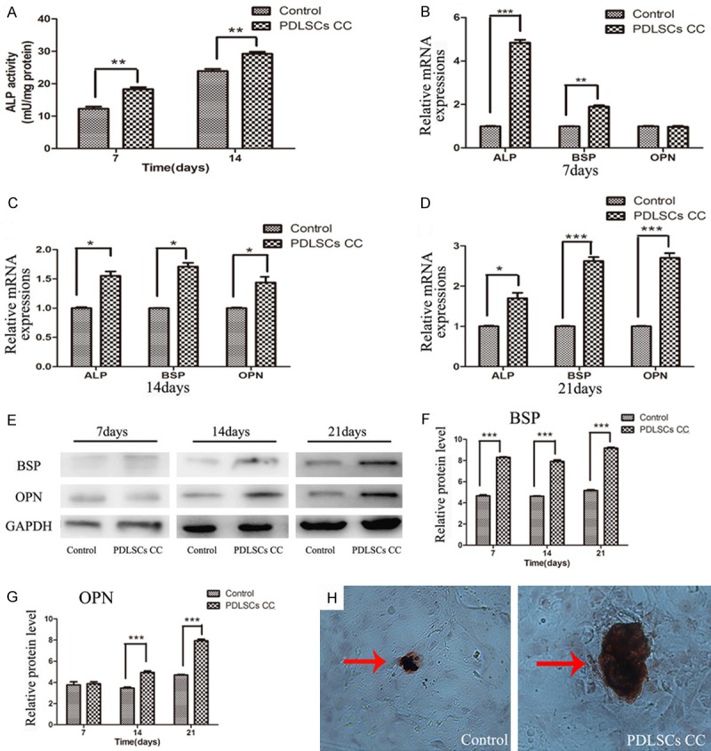
Figure 2.

Effects of PDLSCs on the osteogenenesis in MC3T3-E1 cells in the co-culture system. A. ALP activity in MC3T3-E1 cells co-cultured with PDLSCs. ALP activity increased in the presence of PDLSCs at day 7 and day 14. B-D. Expressions of ALP, BSP, and OPN genes in MC3T3-E1 cells co-cultured with PDLSCs analyzed by real time-PCR. B. Gene expression after 7 days of induction. C. Gene expression after 14 days of induction. D. Gene expression after 21 days of induction. E. Time-related expression of BSP and OPN in MC3T3-E1 cells analyzed by Western blot. F. Quantitative analysis of the protein expression of BSP. G. Quantitative analysis of the protein expression of OPN. H. Mineral matrix deposition (Alizarin red staining) when MC3T3-E1 cells were co-cultured with PDLSCs for 21 days (original magnification 200×). *P < 0.05; **P < 0.01; ***P < 0.001 vs. control group.
