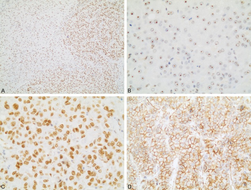
Figure 1.

Representative immunohistochemical staining of NAT10 protein in HCC and peritumoral tissues. A. Immunostaining pattern of NAT10 in tumor cells was more intense than nonneoplastic cells. B. Peritumoral tissues showed only nucleolar staining. C. HCC tissues showed strong nuclear staining. D. High NAT10 expression in membranous of poorly-differentiated tumor cells (A. 100×, B, C. 400×, D. 200× magnification).
