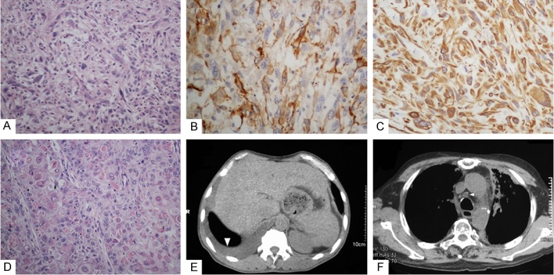
Figure 1.

A. The portion of spindle-shaped tumor cells. B. Spindle-shaped cells were positive for smooth muscle actin. C. Spindle-shaped cells were positive for vimentin. D. The portion of SqCC with frequently keratin pearls. E. CT scan revealed right pleural effusion, laboratory examination found no tumor cells. F. CT scan shows the enlargement of mediastinal lymph node.
