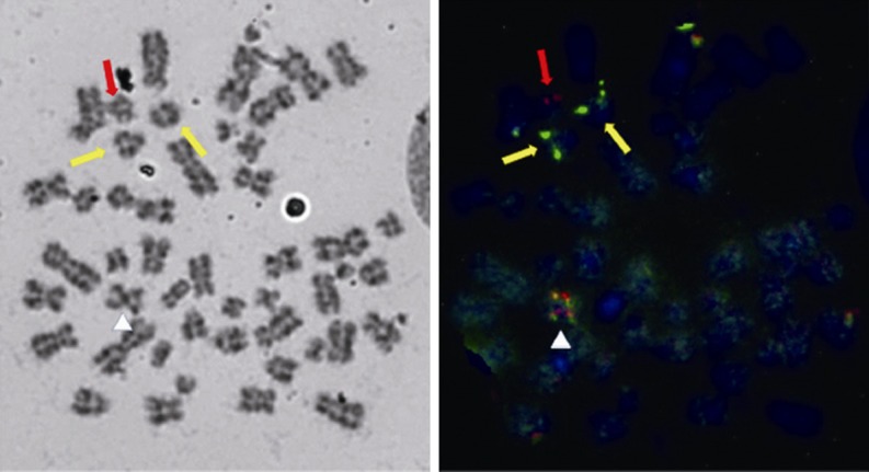
Figure 6.

The FISH analysis at second relapse. The metaphase FISH on bone marrow cytogenetic specimens detected 2 green PML signal on both normal chromosome 15 (yellow arrows), 1 red RAR signal on the normal chromosome 17 (red arrow) and 2 red/green fusion signals on both the long arms of isochromosome 17 (white triangle symbol). The FISH results confirmed the cryptic insertion of PML on chromosome 15 to isochromosome 17 in this case.
