
Figure 1. Intracellular Ca2+ responses in carotid body type I versus type II cells during chemostimulation, and evidence for crosstalk .
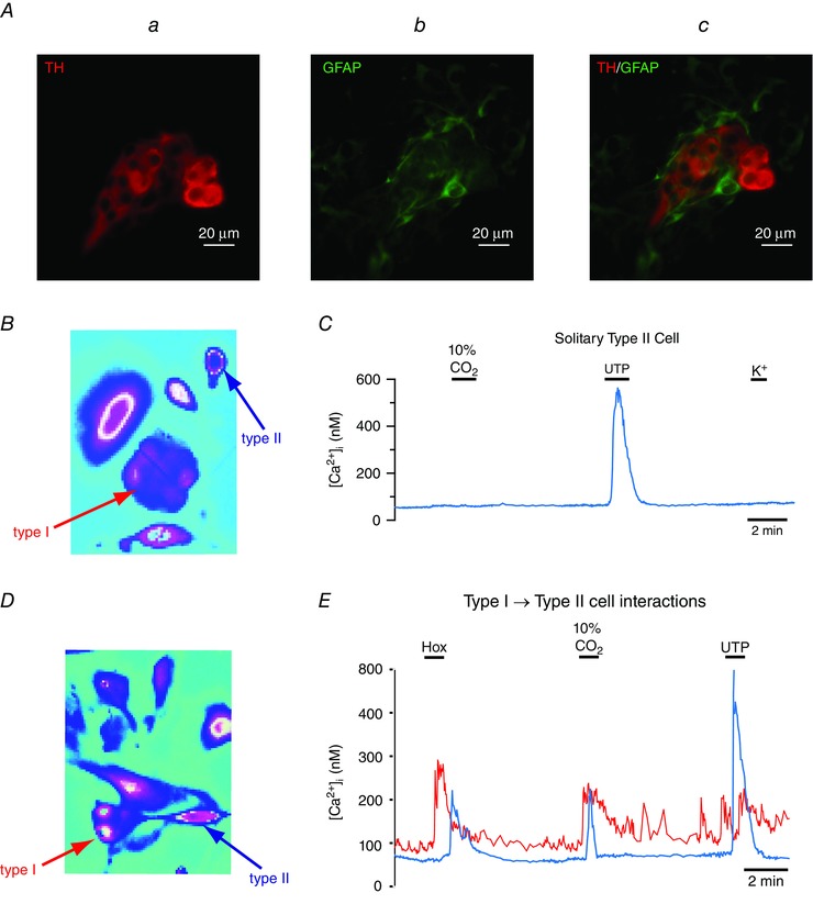
A, immunofluorescence staining of rat carotid body culture showing typical chemoreceptor cluster containing tyrosine hydroxylase (TH)‐positive type I cells (red; Aa), contiguous GFAP‐positive type II cells/processes (green; Ab), and merge (Ac). ‘Solitary’ type II cell (blue arrow), well isolated from a type I cluster (red arrow) in B, fails to respond directly to the chemostimulus isohydric hypercapnia (10% CO2; pH = 7.4) or the depolarizing stimulus high K+, but elicits a robust rise in intracellular Ca2+ to the P2Y2R agonist UTP (C). By contrast, type II cells located near a type I cluster as in D may respond indirectly to chemostimuli such as hypoxia (Hox) ( ∼25 mmHg) and isohydric hypercapnia as shown in E (blue trace); note the delay in type II cell response relative that of a type I cell (E; red trace) in the cluster (D), consistent with crosstalk from type I to type II cells.