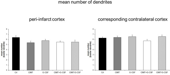
Fig 2. Quantitative analysis of Golgi Cox-impregnated layer V pyramidal neurons in the peri-infarct cortex and in the corresponding contralateral cortex.

There were no differences in the number of basilar dendrites between the various experimental groups neither in the peri-infarct cortex nor in the corresponding contralateral cortex (Values are expressed as mean number of basilar dendrites ± SEM).
