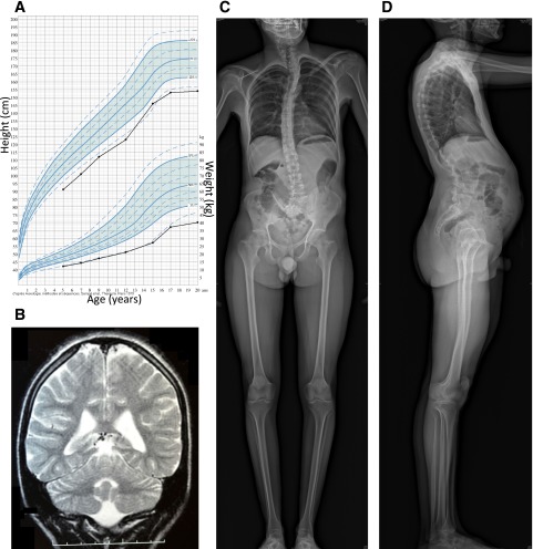
Figure 1.
Imaging of brain and skeleton of patient 1. A: Growth chart, showing growth retardation. B: Coronal T2-weighted brain MRI at age 15 years, showing a moderate white matter rarefaction characterized by increased sulcal size and moderate enlargement of ventricular system. C and D: Skeletal radiographies at age 28 years, showing kyphoscoliosis with tall vertebral bodies and hyperlordosis.

