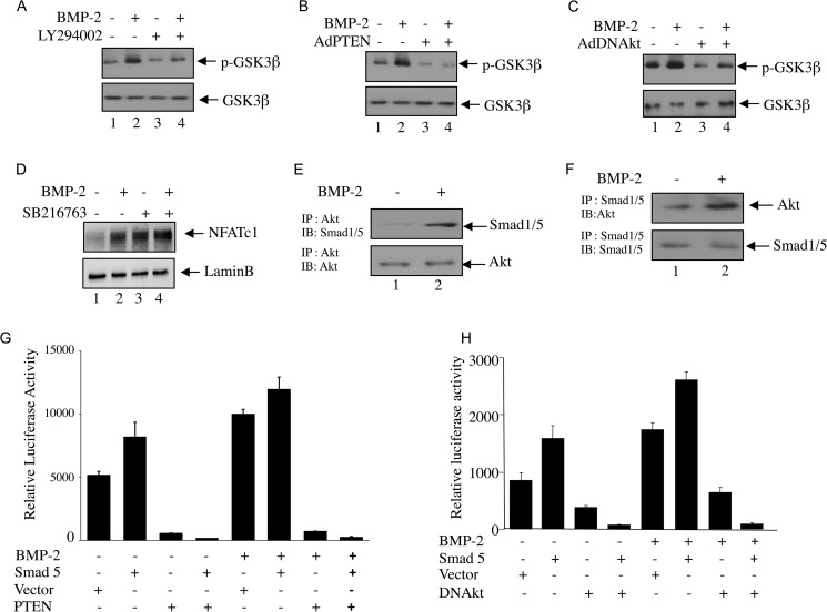
FIGURE 5.
Akt kinase and GSK3β regulate BMP-2-induced NFATc1 expression. A–C, C2C12 cells were treated with Ly294002 (A) or infected with adenovirus expressing PTEN (Ad PTEN; B) or dominant negative Akt kinase (Ad DNAkt; C) before BMP-2 treatment. Cell lysates were immunoblotted with phospho-GSK3β (p-GSK3β) (upper panel) or GSK3β (lower panel) antibody. D, C2C12 cells were treated with 25 μm SB216763 prior to incubation with BMP-2. The nuclear lysates were immunoblotted with NFATc1 (upper panel) or lamin B (lower panel) antibody. E and F, Akt interacts with Smad1/5. Lysates of C2C12 cells treated with BMP-2 were immunoprecipitated (IP) with Akt (E) or Smad1/5 (F) antibody. The immunoprecipitates were immunoblotted (IB) with Smad1/5 (E) or Akt (F) antibody, respectively. G and H, Akt and Smad cooperate for NFATc1 transcription. C2C12 cells were cotransfected with NFATc1-Luc plasmid and empty vector or plasmids expressing Smad5 and PTEN (G) or Smad5 and a dominant negative form of Akt kinase (DNAkt) (H). Cells were incubated with BMP-2, and luciferase activities were measured in the cell lysates as described in Fig. 2. Error bars represent S.E.

