
FIGURE 1.
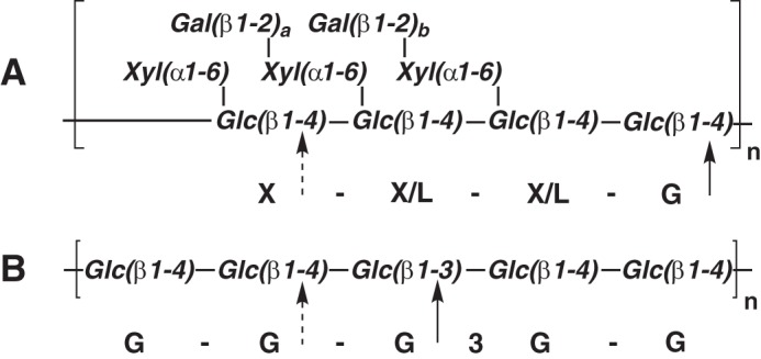
Polysaccharide structures. A, structure of tamarind xyloglucan depicting the repeating Glc4 oligosaccharide moiety and variable galactosylation (a, b = 0 or 1). The primary site of PbGH5A attack (and canonical cleavage site of XyG) is marked with a solid arrow, and the secondary site is marked with a dashed arrow. B, structure of barley β(1,3)/β(1,4)-mixed linkage glucan (depicting GGG3GG). The primary and secondary sites of PbGH5A attack are indicated as for tXyG.