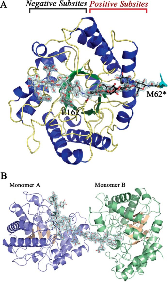
FIGURE 12.

Overall structure of PbGH5A. A, PbGH5A in complex with inhibitor 1. Secondary structure of PbGH5A is shown in schematic representation and color-coded with strands in green, helices in blue, and loops in yellow. Two ligand molecules, XXXG-NHCOCH2Br, are shown in ball-and-stick, in gray and black. The active site positive and negative subsites are indicated. B, asymmetric unit for the PbGH5A(E280A)·XXXGXXXG complex. Secondary structure of PbGH5A is shown in schematic representation and color-coded blue and pink for monomer A and green and pink for monomer B. Two ligand molecules are shown in ball-and-stick, in gray and black.
