
FIGURE 16.
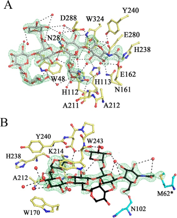
PbGH5A active site in complex with XXXG-NHCOCH2Br. A, negative subsites. The ligand in the active site is in gray, in ball-and-stick, and PbGH5A side chains, involved in binding are in yellow; water molecules are shown as red spheres. Fo − Fc electron density (3.5σ level) is contoured in green around the ligand. B, positive subsites. The ligand is in black, and the residues from adjacent symmetry-related monomer are in cyan. Other coloring is as in A.