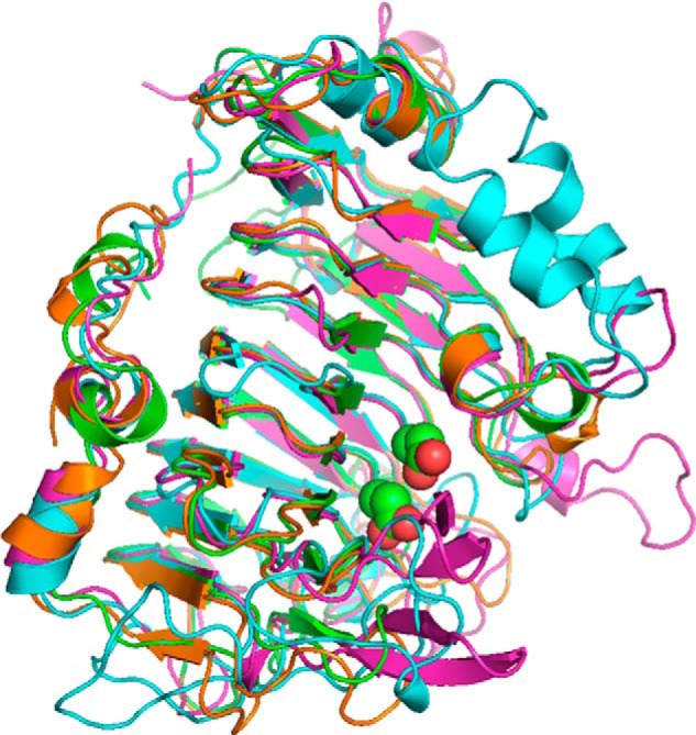
FIGURE 8.

Schematic representation of the superposition of PME structures. The PME2 from A. niger is shown in green, D. carota in orange (PDB code 1gq8), rice weevil (Sitophilus oryzae) in cyan (PDB code 4pmh), and E. chrysanthemi in magenta (PDB codes 2nt6 and 2nt9). The active-site Asp residues are shown as spheres. Relative to Fig. 6A, the view here is rotated ∼90° anti-clockwise about an axis running south-north. The N and C termini of the rice weevil PME are cyan-highlighted; the others are shown in black.
