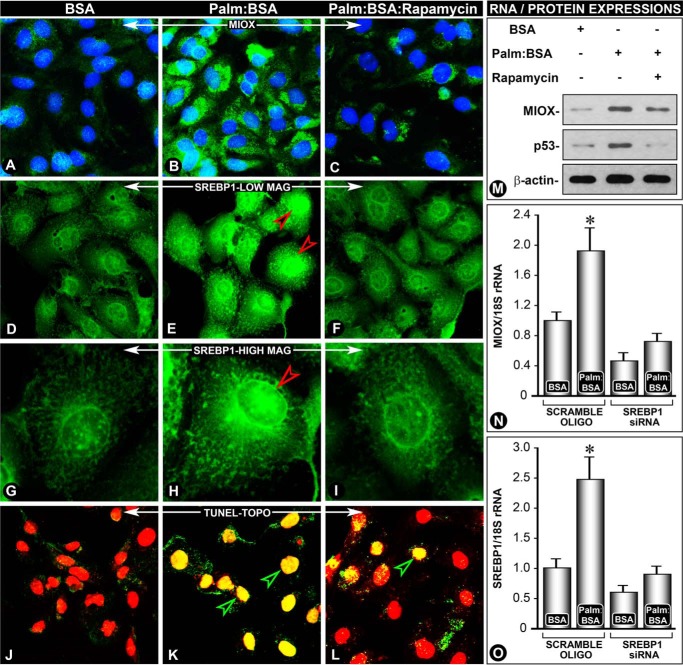
FIGURE 7.
Rapamycin reverses palmitate/BSA-induced Miox, Srebp1, and p53 expression and apoptosis in renal tubular cells. A notable increase in the expression of Miox and Srebp1 was observed following treatment with palmitate/BSA compared with BSA-treated cells alone (B and E versus A and D). The increase was predominantly confined to the nuclear fraction (G and H, arrowhead). The increased expression of Miox and Srebp1 was reduced with rapamycin treatment (C, F, and I), suggesting that the mTOR pathway is involved in the up-regulation of Miox and Srebp. Associated with the increased expression of Miox and Srebp1, a concomitant increase of apoptosis (K, yellow-colored nuclei) was seen, and it was reduced by rapamycin treatment, as assessed by the TUNEL assay (TO-PRO-3 dye) (L, red-colored nuclei). Normally, the intact nuclei yield red fluorescence after staining with this dye (J).The apoptosis observed was most likely mediated by p53, and its increased expression was also reduced by rapamycin (M). A causal relationship between Miox and Srebp1 was established by siRNA experiments where srebp1 siRNA treatment reduced miox as well as srebp1 mRNA expression (N and O). *, p < 0.01 versus control, n = 4.

