FIGURE 8.
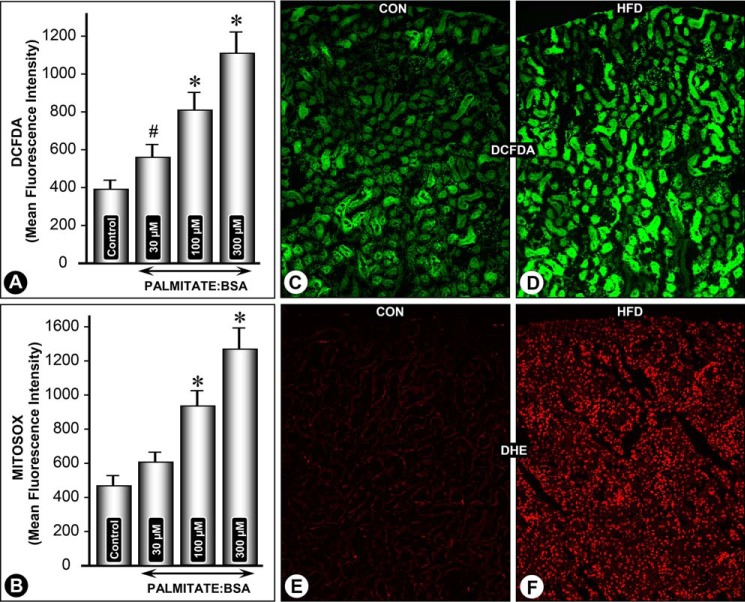
Palmitate/BSA and HFD-induced perturbations in cellular redox in renal tubular cells. A dose-dependent increase in MFI related to CM-H2DCF-DA and MitoSOX staining was observed in HK-2 cells following palmitate/BSA treatment (A and B). Likewise, a notable increase in both CM-H2DCF-DA and DHE staining was observed in kidney sections of mice fed an HFD (D and F versus C and E). DCF staining was mainly confined to the cytoplasm of tubules, although the glomeruli were unaffected. *, p < 0.01 versus control, n = 4.

