
Fig. 3.
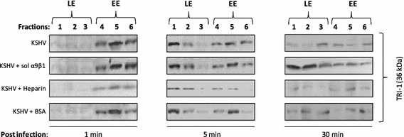
Confirming the presence of KSHV in sucrose gradient fractions at different early time points. Fractions (5 µg protein) collected at different early time points (1, 5, or 30 min) during the course of infection were resolved by SDS-PAGE followed by Western blot analysis with anti-KSHV TRI-1 antibodies