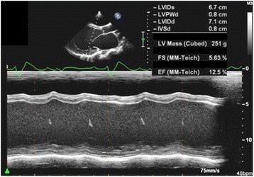
Fig. 1.

M-mode echocardiographic recordings from the index patient showing severe dilatation of the left ventricle (LVIDs: Left ventricle internal diameter during systole; LVIDd: Left ventricle internal diameter in diastole; both are markedly increased) and poor contractility with an ejection fraction (EF) of 12.5 %
