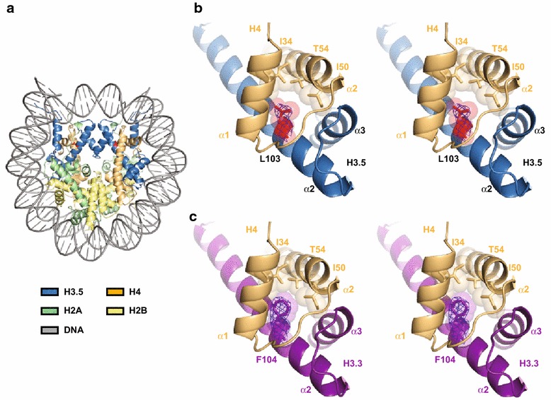
Fig. 2.

Crystal structure of the H3.5 nucleosome. a Overall structure of the H3.5 nucleosome. The H3.5, H4, H2A, H2B, and DNA molecules are colored sky blue, light orange, pale green, pale yellow, and gray, respectively. The H3.5-specific Leu103 residues are colored red, and their side chains are represented. b Stereo view of the H3.5 (sky blue) and H4 (light orange) region around the H3.5 Leu103 residue (red). The 2mFo-DFc electron density map around the H3.5 Leu103 residue is shown as a blue mesh, contoured at 1.5σ. The van der Waals surfaces of the H3.5 Leu103 side chain atoms, and the H4 Ile34, Ile50, and Thr54 side chain atoms, are represented. c Stereo view of the H3.3 (deep purple) and H4 (light orange) region around the H3.3 Phe104 residue in the H3.3 nucleosome structure [PDB:3AV2] [27]. The 2mFo-DFc electron density map around the H3.3 Phe104 residue is shown as a blue mesh, contoured at 1.5σ. The van der Waals surfaces of the H3.3 Phe104 side chain atoms, and the H4 Ile34, Ile50, and Thr54 side chain atoms, are represented
