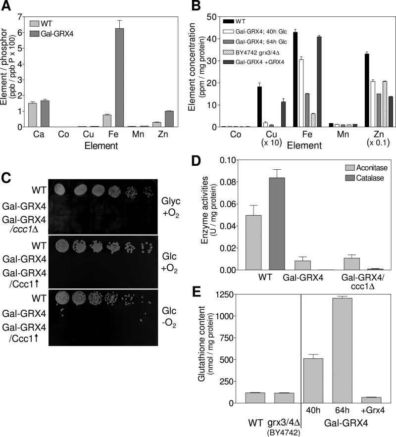
Fig. 3. Deficiency in Grx3/4 results in cytosolic iron and GSH accumulation.
The metal content of (A) wild-type (WT) and Gal-GRX4 cells (depleted for 64 h) and (B) mitochondria isolated from the indicated strains was determined by ICP-MS. (C) The indicated strains lacking (ccc1Δ) or overproducing Ccc1 (Ccc1↑) were cultivated in SD medium for 40 h. Tenfold serial dilutions were spotted onto SC agar plates containing glycerol (Glyc) or glucose (Glc), and cultivated at 30°C under aerobic (+O2) or anaerobic (−O2) conditions. (D) WT, Gal-GRX4 and Gal-GRX4/ccc1Δ cells were grown in SD medium for 64 h, and aconitase and catalase enzyme activities were determined. (E) GSH levels were determined in cell extracts from WT, BY4742 grx3/4Δ, Gal-GRX4 (depleted for 40 h or 64 h) and Gal-GRX4 cells expressing GRX4 from a plasmid (+Grx4).

