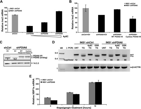
Figure 5.
Degradation of insulin transcripts in the absence of PDIA6 is mediated by IRE1. A) PDIA6-depleted INS1 832/13 cells were treated with different concentrations (10, 30, and 90 μM) of 4μ8C for 8 h. Untreated shCtrl samples served as an internal reference. B) PDIA6-depleted INS1 cells were transiently transfected with empty vector (EV) or wild-type (WT) or cysteine-free V5-tagged PDIA6. Ins2 transcript was measured by qPCR at 48 h after transfection. C) Samples in (B) were immunoblotted to detect the level of exogenous PDIA6 over the endogenous. D) PDIA6-sufficient and -depleted INS1 cells were treated with 500 nM TG for 6 h. In addition to TG, one sample was pretreated with 4μ8C and another with the GSK inhibitor as the negative control. Unspliced (u) and spliced (s) XBP1 mRNAs were amplified by RT-PCR. β-Actin served as control for RNA recovery. The percentage of spliced XBP1 out of the total is reported under each lane. E) PDIA6-sufficient and -depleted INS1 cells were treated with 500 nM TG for the indicated times, and XBP1 splicing was analyzed by qPCR.

