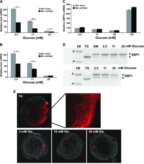
Figure 6.
PDIA6 depletion exacerbates chronic glucose exposure–induced RIDD activity. A, B) PDIA6-sufficient and -depleted INS1 832/13 cells were exposed to the indicated concentrations of glucose for 2 days. Ins2 (A) and Ins1 (B) transcripts were measured by qPCR. Each value was normalized to shCtrl cells in 11 mM glucose. Data are means ± sd of 3 independent experiments. *P ≤ 0.05; **P ≤ 0.01; Student’s t test; shPDIA6 vs. the corresponding shCtrl condition. C) XBP1 splicing was measured from samples as in (A) by qPCR. Cells were also treated with 500 nM TG for 6 h as a positive control for induction of XBP1 splicing. D) INS1 832/13 cells were exposed to the indicated concentrations of glucose for 3 days in 2 independent experiments. Unspliced (u) and spliced (s) XBP1 mRNAs were amplified by RT-PCR. β-Actin served as the control for RNA recovery. E) Mouse islets were transfected with the tdTomato XBP1 splicing reporter and then exposed to different glucose concentrations. TG treatment served as the positive control to identify transfected cells, given that only a portion of islet cells are responsive to ER stress.

