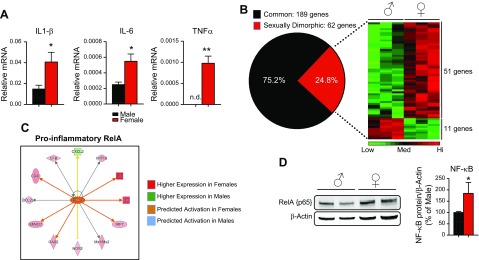
Figure 1.
Hepatic inflammatory genes are expressed in a sexually dimorphic manner. A) qPCR for IL-1β, IL-6, and TNFα from livers of male and female C57BL/6J mice. Data are gene vs. PPIB mRNA levels (means ± sem, n = 3 animals). B) Common and sexually dimorphic genes from nanostring analysis. Heat map of sexually dimorphic genes showing 51 genes have higher expression in female liver and 11 genes have higher expression in males (n = 3 animals). C) Network map of RelA target genes from nanostring data. Males did not show activation of this pathway, hence no blue indicators. D) Western blot analysis of RelA in male and female liver homogenates. RelA protein was normalized to β-actin and expressed as a percentage of that in male liver (n = 4–5 animals). n.d., nondetectable. *P < 0.05, **P < 0.01.

