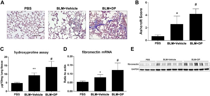
Figure 4.
Exacerbated lung fibrosis in resolvable fibrosis model after DP treatment. Wild-type C57BL/6 female mice (n = 5) were exposed to BLM (2.5 U/kg) by oropharyngeal aspiration. DP (5 mg/kg) and its vehicle control were administered by intraperitoneal injection 2 times per day after 14 d of BLM exposure. Mice were killed at d 21. A) Masson trichrome staining for visualization of collagen deposition (blue). Sections are representative of n = 5 mice from each group. Scale bars, 200 µm. B) Pulmonary fibrosis was evaluated by Ashcroft method. C) Hydroxyproline measurement in lung tissue. D) Fibronectin mRNA expression examined by RT-qPCR. E) Western blot analysis for fibronectin expression in whole lung protein lysate. All data are representative of duplicate repeats of experiments and presented as means ± sem. *P < 0.05, **P < 0.01 for difference between PBS and BLM + vehicle groups; #P < 0.05 for difference between BLM + vehicle and BLM + DP groups.

