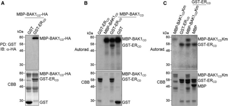
Figure 6. Transphosphorylation between the cytosolic kinase domains of BAK1 and ER.

(A) BAK1CD interacts with ERCD in vitro. MBP-BAK1CD-HA proteins were incubated with GST or GST-ERCD glutathione beads, and the pull-down (PD) proteins were immunoblotted with α-HA antibody (top panel). The CBB staining of input proteins is shown on the bottom panel. (B) The phosphorylation of ERCD by BAK1CD (top panel). (C) The phosphorylation of BAK1CD by ERCD (top panel). The kinase assays were performed using ERCD and BAK1CD kinase mutant (BAK1CDKm) proteins as substrates in (B) and (C) respectively. The CBB staining of input proteins is shown on the bottom panels. The experiments were repeated three times with similar results. (see also Figure S6B).
