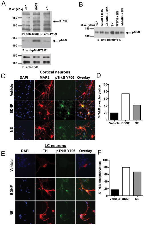
Fig. 4. NE triggers TrkB phosphorylation in cultured cortical and locus coeruleus neurons.
(A) Primary cortical neurons were treated with vehicle, BDNF (100 ng/ml), or NE (500 nM) for 15 min. Total TrkB was immunoprecipitated with anti-TrkB antibody, followed by immunoblotting analysis with anti-PY99 antibody (top panel). Total lysates were also analyzed with anti-pTrkB 817 antibody (middle panel) and anti-TrkB antibody (bottom panel). (B) Primary cortical neurons from TrkB F616A knock-in mice were incubated with vehicle, K252a (100 nM), or 1NMPPI (100 nM) for 2 h and then treated with vehicle or NE (500 nM) for 15 min, followed by immunoblotting analysis with anti-pTrkB 817 antibody. Primary cortical (C, D) and LC (E, F) neurons were pretreated with vehicle, BDNF (100 ng/ml) or NE (500 nM) for 15 min. Neurons were fixed and costained with the nuclear marker DAPI (blue), the neuronal marker MAP2, or the catecholaminergic marker TH (red), and anti-pTrkB 706 (green). Shown are representative immunofluorescent images (C, E) and % of the 150–200 total neurons counted for each condition that were also pTrkB-positive (D, F).

