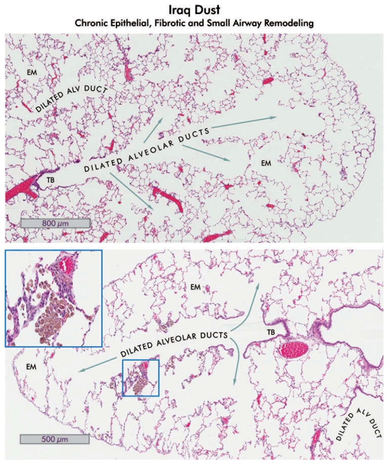
FIGURE 10.
Alveolar duct dilation, emphysema, and Iraq dust in lungs of rats which received 10 mg/kg bw Ir8 dust, 150 d after exposure. Hematoxylin and eosin-stained sections of rat lung are shown. Upper: The lung shows dilation of alveolar ducts and emphysema (EM). No lesions of pneumoconiosis are present, indicating total clearance of the dust in this region. The terminal bronchiole is indicated (TB; 4×). Lower: The central bronchiole with small airways extending nearly to the pleura are shown in the lung of a different rat. The bronchiole and alveolar ducts are dilated, and small areas of emphysema (EM) are evident (4×). There are small clusters of pigmented macrophages containing dust in some of the alveoli (inset, 40×). The epithelium adjacent to the macrophages is slightly hyperplastic and the interstitium is mildly thickened. Compare with the normal lung shown in Figure 3.

