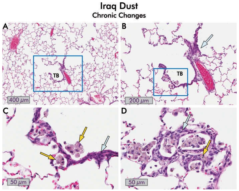
FIGURE 11.
Chronic changes following exposure to Iraq dust. (A) Hematoxylin and eosin stained section of a rat lung 150 d after exposure to 10 mg/kg bw Iraq dust. The terminal bronchiole (TB) in this section appears normal although the associated alveolar ducts are slightly dilated, and there is mild emphysema. There is minimal inflammation and fibrosis (2×). (B) Close-up of boxed area in (A). The terminal bronchiole appears normal, and there is no extension of the bronchiolar epithelium into the alveoli. There is mild interstitial inflammation (blue arrow) and focal accumulation of macrophages in the boxed area (8×). (C) Higher magnification view of the boxed area in (B). There are a few alveolar macrophages containing dust (yellow arrows), and mild alveolar hyperplasia of the epithelium adjacent to the macrophages (yellow arrows), but no evidence of atypia (40×). (D) Alveolar changes in the lung of a different rat 150 d after exposure to Iraq dust. Only very occasional accumulations of dust in macrophages were evident in this animal. The epithelial changes are hyperplastic with minimal dysplasia (blue arrow). There is mild chronic inflammation (yellow arrow) but no evidence of fibrosis (40×).

