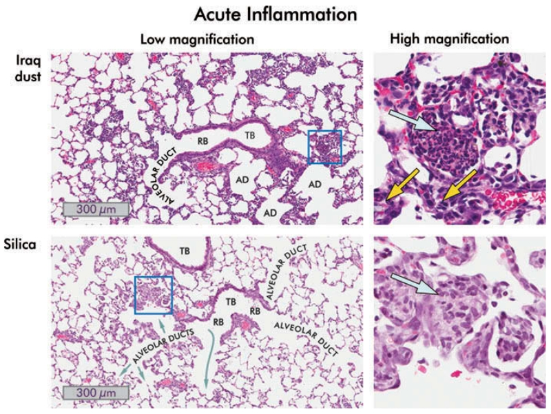
FIGURE 5.
Inflammation in the lungs of rats one day following instillation of 10 mg/kg bw Iraq or silica dust. Low- (8×) and high-magnification (40×) views of hematoxylin and eosin stained lung sections are shown. Iraq dust: A terminal bronchiole (TB), a respiratory bronchiole (RB), and alveolar ducts (AD) are shown. Many alveoli adjacent to the bronchiole are filled with inflammatory cells, predominately neutrophils and eosinophils. In the higher magnification view of the boxed area, multinucleated polymorphonuclear leukocytes and eosinophils are present (blue arrow), and acute inflammatory cells are visible in the interstitium (yellow arrows). Silica: The low-magnification image contains two small terminal bronchioles together with alveolar ducts. The alveoli are filled with neutrophils and, predominately, macrophages. The high-magnification view of the boxed area shows the intra-alveolar exudate in which macrophages predominate (blue arrow). The epithelium lining the alveoli is hyperplastic.

