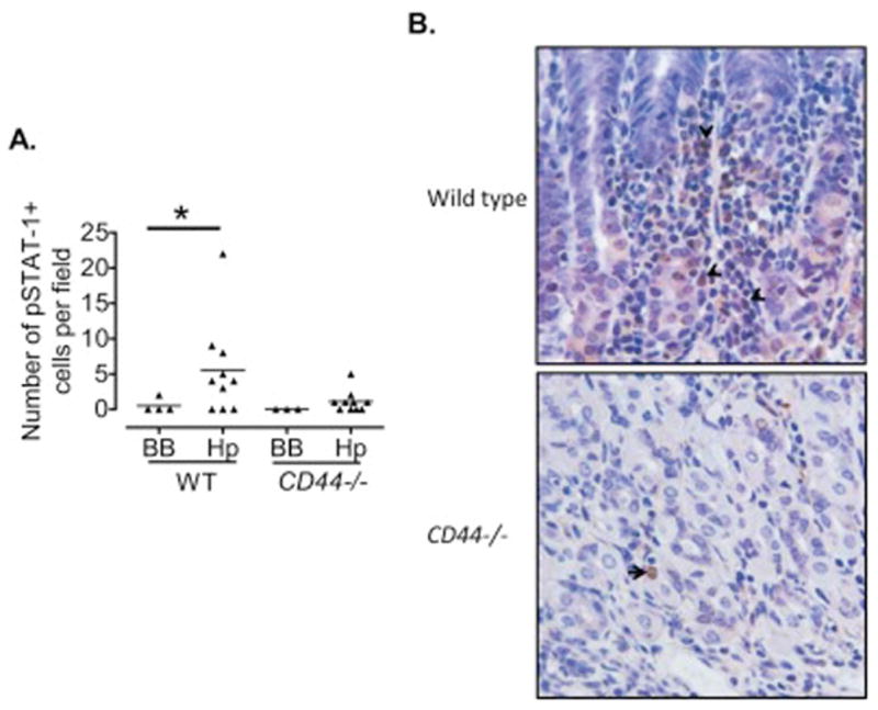
Figure 4. Reduced expression of pSTAT-1 in CD44−/− mice following H. pylori infection.

A. The number of pSTAT-1+ cells in 10 high power fields (200X) was assessed using immunohistochemistry in the gastric mucosae of CD44−/− and wild type mice infected with H. pylori for 7 months. B. Representative immunohistochemistry showing pSTAT-1 positive cells in the gastric mucosa (200X magnification). *p=0.07; BB, Brucella broth.
