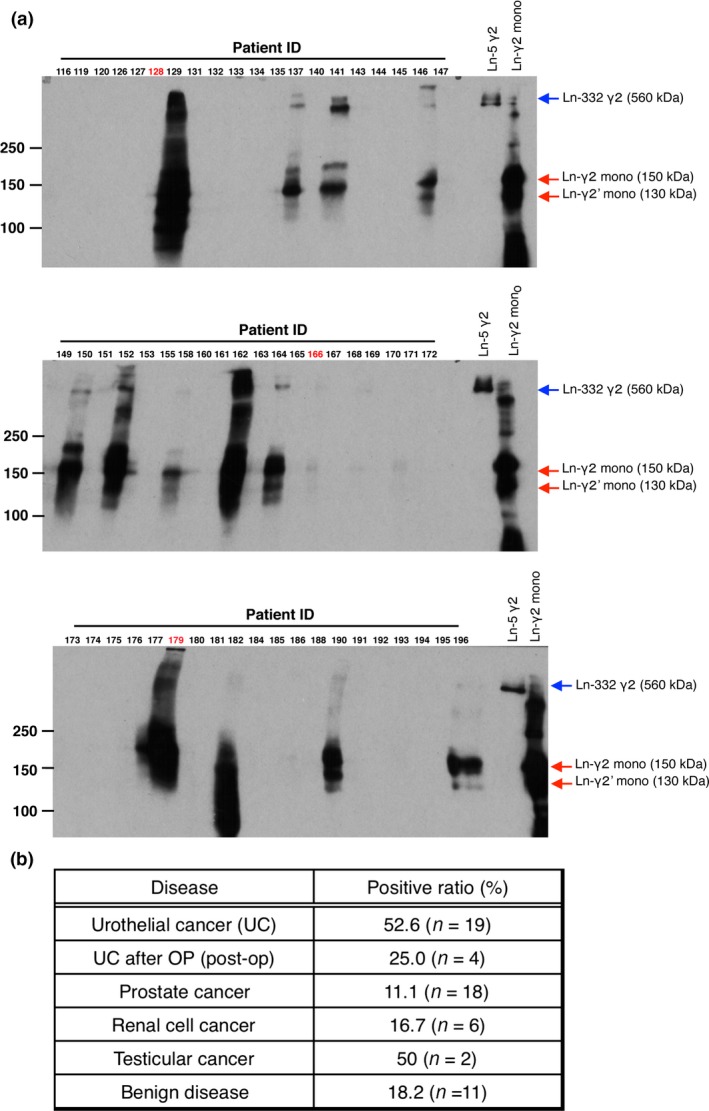
Figure 1.

Detection of laminin‐γ2 (Ln‐γ2) in urine from urothelial cancer patients by Western blotting. (A) Sixty urine samples (80 μL) were concentrated using ice‐cold acetone and were subjected to Western blotting using D4B5 mAb under non‐reducing conditions. Ln‐332 and monomeric Ln‐γ2 (Ln‐γ2 mono) were used as positive controls. Red arrows indicate monomeric Ln‐γ2 and its oligomeric forms; blue arrows indicate Ln‐332γ2. Urothelial cancer tissues (red numbers) were subsequently analyzed using immunohistochemistry. (B) Summary of the Ln‐γ2‐positive urine ratio (%) from patients with malignant and benign urologic disease.
