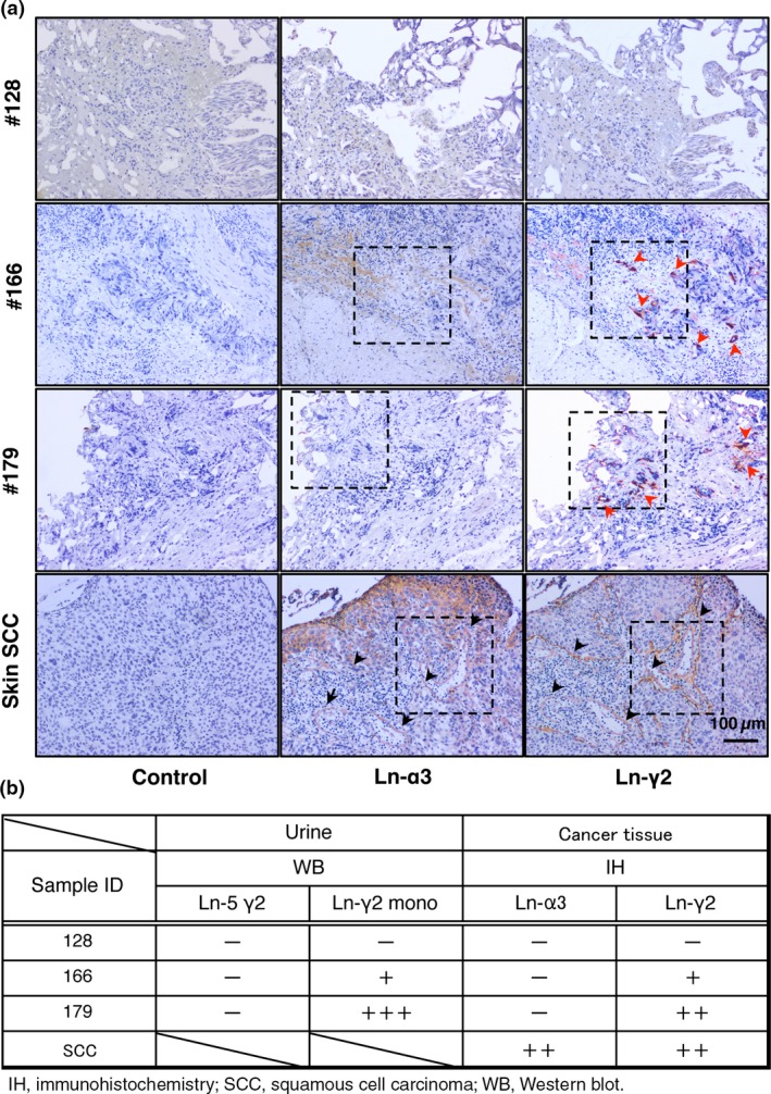
Figure 2.

Detection of laminin (Ln‐α3 or ‐γ2) in urothelial cancer (UC) by immunohistochemistry (IH). (A) IH analysis of Ln‐α3 and ‐γ2 (components of Ln‐332). Red arrowheads indicate muscle invasive UC tissue expressing Ln‐γ2 in patients 166 and 179, but not in patient 128 (non‐muscle invasive UC). Black arrowheads indicate basement membranes expressing Ln‐α3 and ‐γ2 in skin squamous cell carcinoma (SCC). Representative views of specimens are indicated with dashed lines. (B) Ln‐α3 and ‐γ2 expression in UC specimens. WB, Western blot.
