Figure 3.

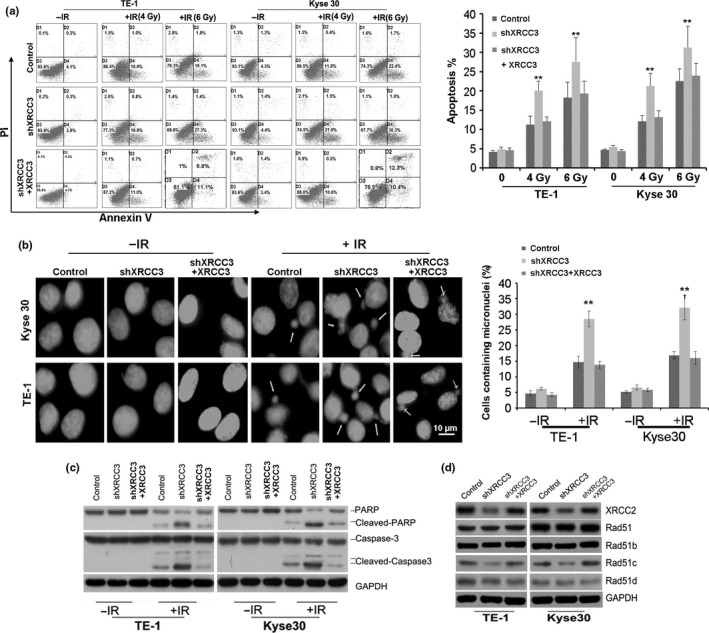
Silencing of XRCC3 promotes ionizing radiation (IR)‐induced apoptosis and mitotic catastrophe (MC) in esophageal squamous cell carcinoma (ESCC) cells. (a) Silencing of XRCC3 promoted IR‐induced apoptosis in both Kyse30 and TE‐1 cells. Annexin V and propidium iodide staining was used to determine the percentage of cells undergoing apoptosis. The percentage of cells with apoptosis is also shown (right). Data represent mean values and SE (*P < 0.05; **P < 0.01, Student's t‐test). (b) Inhibition of XRCC3 increased micronuclei in both Kyse30 and TE‐1 cells exposed to 6 Gy IR. Cells were stained with DAPI and examined for nuclear morphology. Arrows indicate micronuclei in interphase (left). The percentage of cells with micronuclei is also shown (right). Data represent mean values and SE. (Bar equals 10 um. *P < 0.05; **P < 0.01, Student's t‐test.) (c) Silencing of XRCC3 increased the levels of cleaved PARP and cleaved caspase‐3 in both TE‐1 and Kyse30 cells exposed to 6 Gy IR. (d) Inhibiton of XRCC3 in ESCC cells downregulated the protein expression of XRCC2 and Rad51c. However, XRCC3 knockdown did not alter the expression of Rad51, Rad51b or Rad51c. GAPDH was used as a loading control. After restoration of XRCC3, the altered cell apoptosis, mitotic catastrophe (MC), protein expression of XRCC2 and RAD51C were all recovered. All data are derived from three individual experiments.
