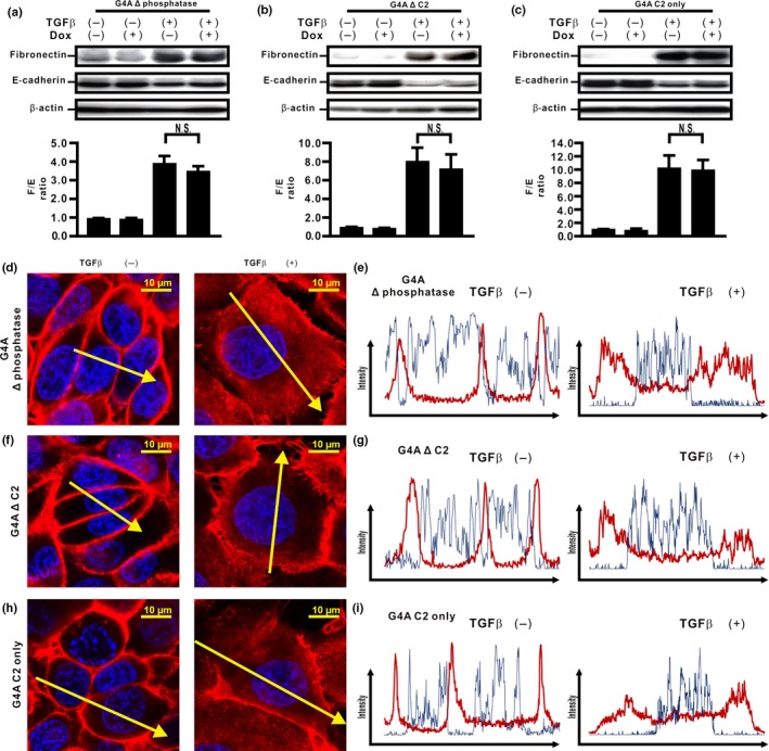
Figure 3.

Phosphatase and C2 domains of unphosphorylated PTEN are essential for inhibition of transforming growth factor β (TGFβ)‐induced epithelial–mesenchymal transition in H358 cells. (a–c) Fibronectin (F) and E‐cadherin (E) were analyzed by Western blotting. The F/E ratio is shown in comparison to that in cells treated with vehicle in the absence of Dox. A representative blot from three independent experiments is shown. Data shown represent the means ± SE. The experiment was repeated three times with similar results. (d–i) The fluorescence intensity of β‐catenin was evaluated. Left and right images in (d,f,h) show cells without and with TGFβ stimulation, respectively. Upper and lower panels in (e,g,i) plot the fluorescence intensity of β‐catenin (red) and nucleus (blue) over a cross‐section of cells without and with TGFβ stimulation along the selected yellow arrows in (d, f, h), respectively. Data shown are representative of at least three independent experiments.
