
Figure 2.
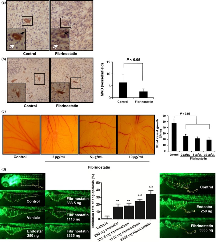
Fibrinostatin inhibits angiogenesis in vivo. (a) Tumor sections from BGC‐823 xenografts were immunostained with anti‐CD31 antibody. Black arrowheads point to the stained vessels. White arrowheads point to tumor cells (left) and necrotic tumor cells (right). Scale bar = 50 μm (b) Tumor sections from BGC‐823 xenografts immunostained with anti‐CD31 antibody were used to examine the vessel number. (×200 magnification, left). Boxed regions show CD31‐positive structures magnified at ×400 in the insets. Mean ± SD microvascular density (MVD) per field was calculated (right). P < 0.05 (unpaired Student's t‐test). Scale bar = 100 μm. (c) Fibrinostatin inhibited angiogenesis in chick chorioallantoic membrane (CAM). (d) Fibrinostatin inhibited angiogenesis in transgenic zebrafish. *** P < 0.001; **P < 0.01; (Student's t‐test).