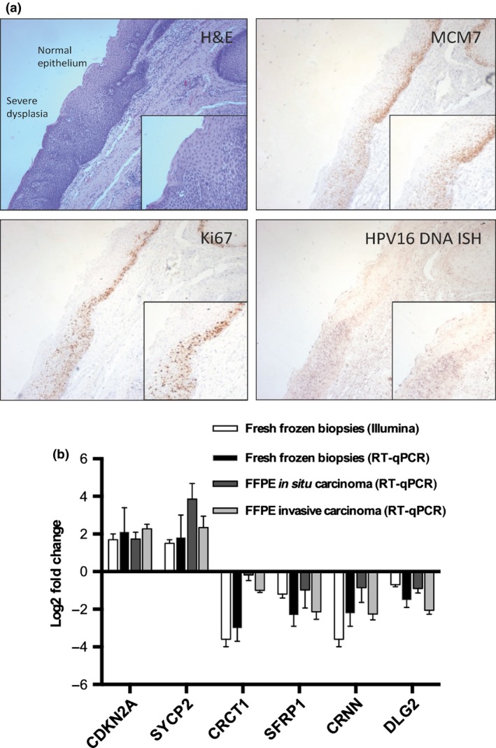
Figure 3.

(a) H&E staining showing the junction between normal epithelium and severe dysplasia in human papillomavirus (HPV)‐positive tonsil squa‐mous cell carcinoma (top left). Although some flattening of cells at the apical surface may exist, the majority of the abnormal epithelium has a poorly differentiated dense basaloid appearance with nuclear pleomorphism. MCM7 (top right) and Ki67 (bottom left) staining are present in more than two‐thirds of the abnormal epithelium. In situ hybridization reveals an elevated level of HPV 16 DNA (bottom right) in the dysplastic epithelium when compared to the normal region (magnification, ×100; inset, ×150; Infinity capture software). ISH, in situ hybridization. (b) Reverse transcription–real‐time quantitative PCR (RT‐qPCR) analysis for all HPV‐positive sample types showing significant differential expression of SYCP2 (log2 fold change, 3.1 [95% confidence interval, 1.8–4.4]; P < 0.01) in premalig‐nant (in situ) tissue.
