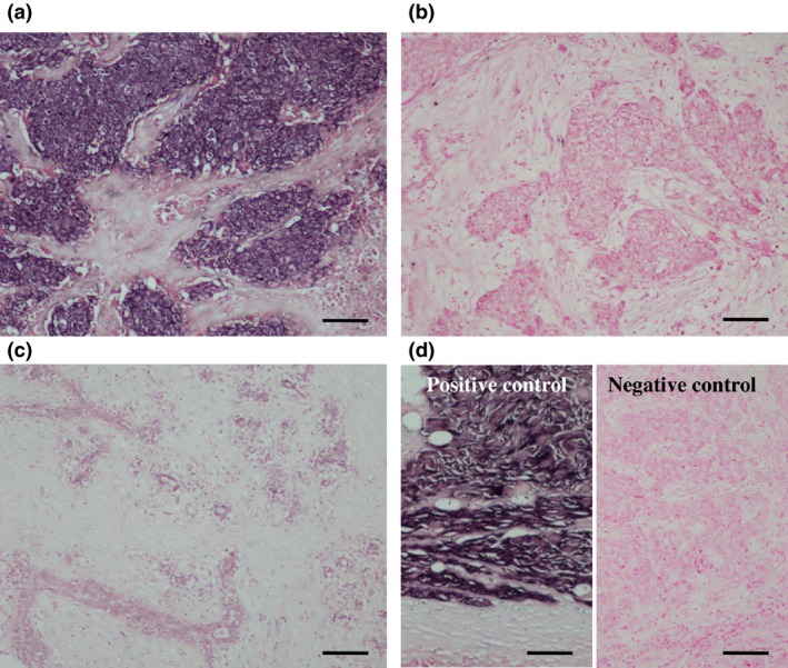
Figure 3.

In situ hybridization (ISH) for miR‐1 in breast carcinoma. (a) miR‐1 was localized in the cytoplasm of carcinoma cells. (b) miR‐1‐negative breast carcinoma. (c) Hybridization signal for miR‐1 was focally and weakly detected in the morphologically normal mammary epithelium. (d) Positive control (skeletal muscle tissue; left panel) and negative control (scrambled negative control probe in breast carcinoma; right panel) for miR‐1 ISH. Bar = 100 μm, respectively.
